. 2011 Dec 2:12:52.
doi: 10.1186/1471-2121-12-52.

Analysis of the role of Ser1/Ser2/Thr9 phosphorylation on myosin II assembly and function in live cells

Affiliations

Analysis of the role of Ser1/Ser2/Thr9 phosphorylation on myosin II assembly and function in live cells

Jordan R Beach et al. BMC Cell Biol. .

Erratum in

  • BMC Cell Biol. 2012;13:11

Abstract

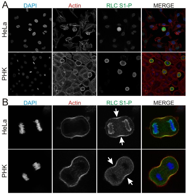
Background: Phosphorylation of non-muscle myosin II regulatory light chain (RLC) at Thr18/Ser19 is well established as a key regulatory event that controls myosin II assembly and activation, both in vitro and in living cells. RLC can also be phosphorylated at Ser1/Ser2/Thr9 by protein kinase C (PKC). Biophysical studies show that phosphorylation at these sites leads to an increase in the Km of myosin light chain kinase (MLCK) for RLC, thereby indirectly inhibiting myosin II activity. Despite unequivocal evidence that PKC phosphorylation at Ser1/Ser2/Thr9 can regulate myosin II function in vitro, there is little evidence that this mechanism regulates myosin II function in live cells.

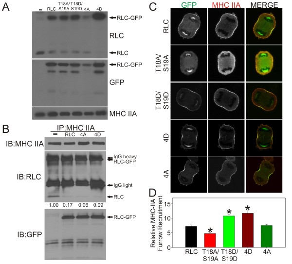
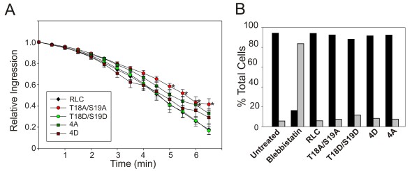
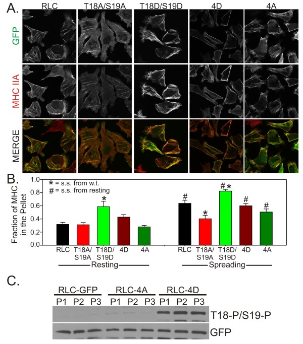
Results: The purpose of these studies was to investigate the role of Ser1/Ser2/Thr9 phosphorylation in live cells. To do this we utilized phospho-specific antibodies and created GFP-tagged RLC reporters with phosphomimetic aspartic acid substitutions or unphosphorylatable alanine substitutions at the putative inhibitory sites or the previously characterized activation sites. Cell lines stably expressing the RLC-GFP constructs were assayed for myosin recruitment during cell division, the ability to complete cell division, and myosin assembly levels under resting or spreading conditions. Our data shows that manipulation of the activation sites (Thr18/Ser19) significantly alters myosin II function in a number of these assays while manipulation of the putative inhibitory sites (Ser1/Ser2/Thr9) does not.

Conclusions: These studies suggest that inhibitory phosphorylation of RLC is not a substantial regulatory mechanism, although we cannot rule out its role in other cellular processes or perhaps other types of cells or tissues in vivo.


Figures

Figure 1
Figure 1
RLC Ser1 phosphorylation is elevated in mitotic cells and localizes to the contractile ring. (A) HeLa and primary human keratinocytes (PHK) under normal growth conditions were fixed and immunostained for RLC Ser1-P (green) and stained for actin (red). (B) RLC Ser1-P is specifically enhanced in the contractile ring of dividing cells.
Figure 2
Figure 2
Expression, localization and assembly of MHC IIA during cell division. (A) Whole cell lysates of HeLa cells expressing the indicated RLC construct were probed for total RLC, GFP and MHC IIA. (B) MHC IIA was immunoprecipitated from the indicated cell line and the eluate was probed for MHC IIA, RLC and GFP. Note that the RLC-GFP and IgG heavy chain migrate too close to differentiate. The number under each construct indicates the relative amount of RLC present after normalization to MHC IIA based on densitometry. (C) Anaphase cells expressing the indicated RLC construct (green) were fixed and stained for MHC IIA (red). Representative images are shown. (C) MHC IIA recruitment was quantitated for a minimum of 13 cells. Data are mean +/- SEM. * indicates p = < 0.003.
Figure 3
Figure 3
Analysis of furrow ingression and multinucleation. (A) HeLa cells expressing the indicated RLC construct were imaged throughout cell division. Furrow width was measured every 30 seconds for a minimum of 5 cells. Data are mean +/- SEM. * indicates p = <0.05. (B) HeLa cells expressing the indicated RLC construct or treated with 60 uM blebbistatin for 24 hours were analyzed for nucleation state using propidium iodide and flow cytometry. Black bars represent cells with DNA content of less than 4n. Grey bars represent cells with DNA content of 4n or greater.
Figure 4
Figure 4
MHC IIA assembly in resting and spreading cells. (A) HeLa cells in normal growth conditions expressing the indicated RLC-GFP construct (green) were fixed and immunostained for MHC IIA (red). (B) Triton-insoluble fractionation was performed on HeLa cells in normal growth conditions ("resting") or actively spreading for 1 hr. Data are mean +/- SEM. N ≥ 6 for resting and N ≥ 9 for spreading. # indicates p = < 0.05 relative to the same cells in resting conditions. * indicates p = < 0.05 relative to the wild type RLC in the same conditions. (C) Multiple pellet (P) fractions from the triton-insoluble fractionation studies were assayed for their Thr18/Ser19 phosphorylation levels. GFP was used to show that each pellet contains similar amounts of the indicated RLC-GFP construct.

References

    1. Vicente-Manzanares M, Ma X, Adelstein RS, Horwitz AR. Non-muscle myosin II takes centre stage in cell adhesion and migration. Nat Rev Mol Cell Biol. 2009;10:778–790. doi: 10.1038/nrm2786. - DOI - PMC - PubMed
    1. Adelstein RS, Conti MA. Phosphorylation of platelet myosin increases actin-activated myosin. ATPase activity Nature. 1975;256:597–598. - PubMed
    1. Scholey JM, Taylor KA, Kendrick-Jones J. Regulation of non-muscle myosin assembly by calmodulin-dependent light chain kinase. Nature. 1980;287:233–235. doi: 10.1038/287233a0. - DOI - PubMed
    1. Ikebe M, Hartshorne DJ, Elzinga M. Phosphorylation of the 20,000-dalton light chain of smooth muscle myosin by the calcium-activated, phospholipid-dependent protein kinase. Phosphorylation sites and effects of phosphorylation. J Biol Chem. 1987;262:9569–9573. - PubMed
    1. Nishikawa M, Tanaka T, Hidaka H. Ca2+-calmodulin-dependent phosphorylation and platelet secretion. Nature. 1980;287:863–865. doi: 10.1038/287863a0. - DOI - PubMed

Publication types

LinkOut - more resources