

Application of layered poly (L-lactic acid) cell free scaffold in a rabbit rotator cuff defect model
- PMID: 22136125
- PMCID: PMC3245426
- DOI: 10.1186/1758-2555-3-29
Application of layered poly (L-lactic acid) cell free scaffold in a rabbit rotator cuff defect model
Abstract
Background: This study evaluated the application of a layered cell free poly (L-lactic acid) (PLLA) scaffold to regenerate an infraspinatus tendon defect in a rabbit model. We hypothesized that PLLA scaffold without cultivated cells would lead to regeneration of tissue with mechanical properties similar to reattached infraspinatus without tendon defects.
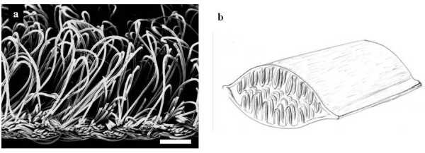
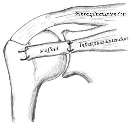
Methods: Layered PLLA fabric with a smooth surface on one side and a pile-finished surface on the other side was used. Novel form of layered PLLA scaffold was created by superimposing 2 PLLA fabrics. Defects of the infraspinatus tendon were created in 32 rabbits and the PLLA scaffolds were transplanted, four rabbits were used as normal control. Contralateral infraspinatus tendons were reattached to humeral head without scaffold implantation. Histological and mechanical evaluations were performed at 4, 8, and 16 weeks after operation.
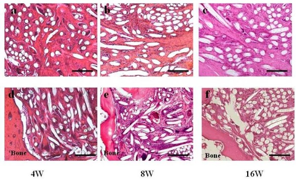
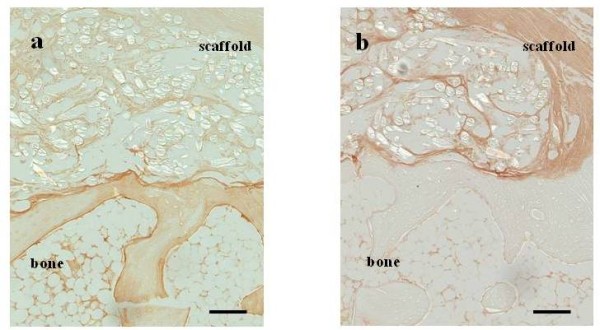
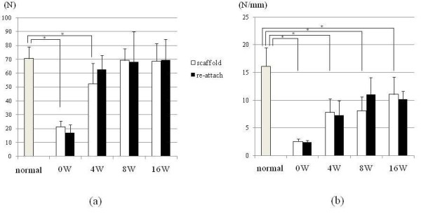
Results: At 4 weeks postoperatively, cell migration was observed in the interstice of the PLLA fibers. Regenerated tissue was directly connected to the bone composed mainly of type III collagen, at 16 weeks postoperatively. The ultimate failure load increased in a time-dependent manner and no statistical difference was seen between normal infraspinatus tendon and scaffold group at 8 and 16 weeks postoperatively. There were no differences between scaffold group and reattach group at each time of point. The stiffness did not improve significantly in both groups.
Conclusions: A novel form of layered PLLA scaffold has the potential to induce cell migration into the scaffold and to bridge the tendon defect with mechanical properties similar to reattached infraspinatus tendon model.
Figures
Similar articles
-
Regeneration of rotator cuff tear using electrospun poly(d,l-Lactide-Co-Glycolide) scaffolds in a rabbit model.Arthroscopy. 2012 Dec;28(12):1790-9. doi: 10.1016/j.arthro.2012.05.887. Epub 2012 Oct 9. Arthroscopy. 2012. PMID: 23058811
-
Potency of double-layered poly L-lactic acid scaffold in tissue engineering of tendon tissue.Int Orthop. 2010 Dec;34(8):1327-32. doi: 10.1007/s00264-009-0917-8. Epub 2009 Dec 5. Int Orthop. 2010. PMID: 19960193 Free PMC article.
-
Ligament regeneration using an absorbable stent-shaped poly-L-lactic acid scaffold in a rabbit model.Int Orthop. 2012 Nov;36(11):2379-86. doi: 10.1007/s00264-012-1660-0. Epub 2012 Sep 14. Int Orthop. 2012. PMID: 22976595 Free PMC article.
-
Rotator Cuff Repair With Autologous Tenocytes and Biodegradable Collagen Scaffold: A Histological and Biomechanical Study in Sheep.Am J Sports Med. 2020 Feb;48(2):450-459. doi: 10.1177/0363546519892580. Epub 2019 Dec 16. Am J Sports Med. 2020. PMID: 31841352
-
Rotator cuff regeneration using a bioabsorbable material with bone marrow-derived mesenchymal stem cells in a rabbit model.Am J Sports Med. 2012 Jun;40(6):1259-68. doi: 10.1177/0363546512442343. Epub 2012 Apr 6. Am J Sports Med. 2012. PMID: 22491821
Cited by
-
Woven collagen biotextiles enable mechanically functional rotator cuff tendon regeneration during repair of segmental tendon defects in vivo.J Biomed Mater Res B Appl Biomater. 2019 Aug;107(6):1864-1876. doi: 10.1002/jbm.b.34279. Epub 2018 Nov 28. J Biomed Mater Res B Appl Biomater. 2019. PMID: 30485649 Free PMC article.
-
Achilles Tendon Repair by Decellularized and Engineered Xenografts in a Rabbit Model.Stem Cells Int. 2019 Aug 29;2019:5267479. doi: 10.1155/2019/5267479. eCollection 2019. Stem Cells Int. 2019. PMID: 31558905 Free PMC article.
-
Evaluation of patches for rotator cuff repair: A systematic review and meta-analysis based on animal studies.Bioact Mater. 2021 Aug 28;10:474-491. doi: 10.1016/j.bioactmat.2021.08.016. eCollection 2022 Apr. Bioact Mater. 2021. PMID: 34901561 Free PMC article.
-
Functionally Graded, Bone- and Tendon-Like Polyurethane for Rotator Cuff Repair.Adv Funct Mater. 2018 May;28(20):1707107. doi: 10.1002/adfm.201707107. Epub 2018 Mar 30. Adv Funct Mater. 2018. PMID: 29785178 Free PMC article.
-
Textile cell-free scaffolds for in situ tissue engineering applications.J Mater Sci Mater Med. 2016 Mar;27(3):63. doi: 10.1007/s10856-015-5656-3. Epub 2016 Jan 22. J Mater Sci Mater Med. 2016. PMID: 26800694 Free PMC article. Review.
References
-
- Karas SE, Giachello TL. Subscapularis transfer for reconstruction of massive tears of the rotator cuff. J Bone Joint Surg Am. 1996;78:239–245. - PubMed
-
- Aoki M, Miyamoto S, Okamura K, Yamashita T, Ikada Y, Matsuda S. Tensile properties and biological response of poly(L-lactic acid) felt graft: an experimental trial for rotator-cuff reconstruction. J Biomed Mater Res B Appl Biomater. 2004;71:252–259. - PubMed
LinkOut - more resources
Full Text Sources
Other Literature Sources
