. 2011 Dec 2:8:169.
doi: 10.1186/1742-2094-8-169.

Neuropeptide Y inhibits interleukin-1β-induced phagocytosis by microglial cells

Affiliations

Neuropeptide Y inhibits interleukin-1β-induced phagocytosis by microglial cells

Raquel Ferreira et al. J Neuroinflammation. .

Abstract

Background: Neuropeptide Y (NPY) is emerging as a modulator of communication between the brain and the immune system. However, in spite of increasing evidence that supports a role for NPY in the modulation of microglial cell responses to inflammatory conditions, there is no consistent information regarding the action of NPY on microglial phagocytic activity, a vital component of the inflammatory response in brain injury. Taking this into consideration, we sought to assess a potential new role for NPY as a modulator of phagocytosis by microglial cells.

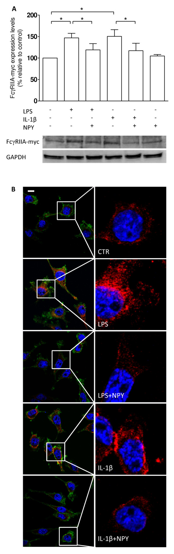
Methods: The N9 murine microglial cell line was used to evaluate the role of NPY in phagocytosis. For that purpose, an IgG-opsonized latex bead assay was performed in the presence of lipopolysaccharide (LPS) and an interleukin-1β (IL-1β) challenge, and upon NPY treatment. A pharmacological approach using NPY receptor agonists and antagonists followed to uncover which NPY receptor was involved. Moreover, western blotting and immunocytochemical studies were performed to evaluate expression of p38 mitogen-activated protein kinase (MAPK) and heat shock protein 27 (HSP27), in an inflammatory context, upon NPY treatment.

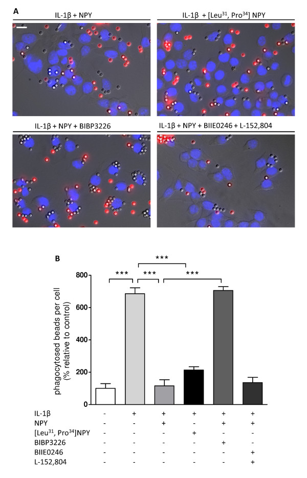
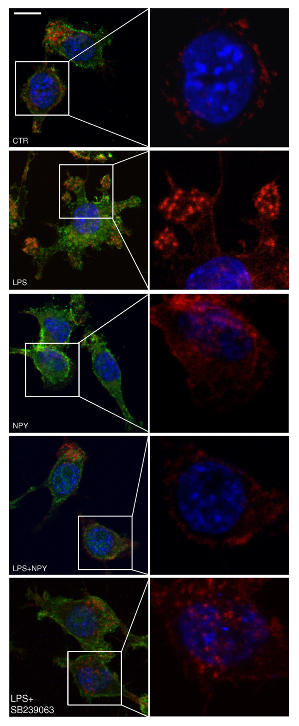
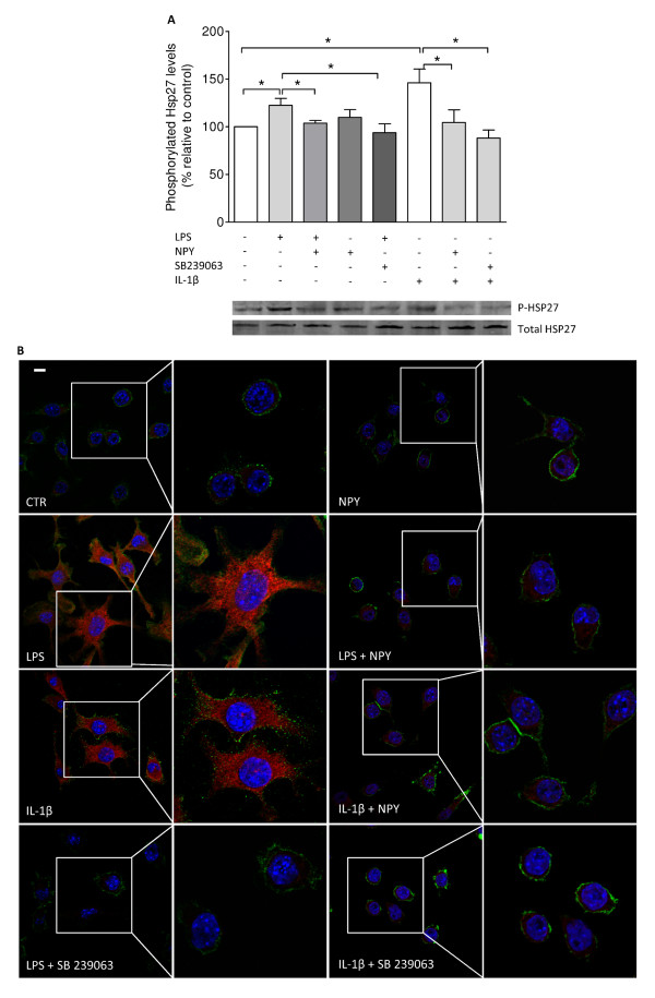
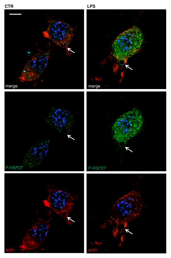
Results: Here, we show that NPY inhibits phagocytosis of opsonized latex beads and inhibits actin cytoskeleton reorganization triggered by LPS stimulation. Co-stimulation of microglia with LPS and adenosine triphosphate also resulted in increased phagocytosis, an effect inhibited by an interleukin-1 receptor antagonist, suggesting involvement of IL-1β signaling. Furthermore, direct application of LPS or IL-1β activated downstream signaling molecules, including p38 MAPK and HSP27, and these effects were inhibited by NPY. Moreover, we also observed that the inhibitory effect of NPY on phagocytosis was mediated via Y1 receptor activation.

Conclusions: Altogether, we have identified a novel role for NPY in the regulation of microglial phagocytic properties, in an inflammatory context.


Figures

Figure 1
Figure 1
NPY inhibits bead phagocytosis by microglial cells. (A) Representative photomicrographs illustrate the inhibitory effect of NPY on LPS-induced phagocytosis. (B) LPS (100 ng/ml) increased bead phagocytosis, while NPY (1 μM) inhibited this effect. Data are expressed as mean ± SEM (n = 3-5) and as a percentage of control (***p < 0.001, using Bonferroni's Multiple Comparison Test). Scale bar 10 μm.
Figure 2
Figure 2
LPS-induced phagocytosis is mediated by IL-1β signaling. (A) Representative photomicrographs illustrate the inhibitory effect of IL-1β, or LPS+ATP alone or with co-treatment with IL-1ra. (B) LPS (100 ng/ml) and ATP (1 mM) co-administration significantly induced bead phagocytosis. This effect was prevented by IL-1ra application (150 ng/ml) suggesting involvement of IL-1β. Direct application of IL-1β (1.5 ng/ml) increased phagocytosis and was completely inhibited by IL-1ra. Data are expressed as mean ± SEM (n = 3-5) and as a percentage of control (***p < 0.001, using Bonferroni's Multiple Comparison Test). Scale bar 10 μm.
Figure 3
Figure 3
NPY inhibits IL-1β-induced phagocytosis via Y1 receptor activation. (A) Representative photomicrographs illustrate the inhibitory effect of NPY acting via Y1 receptor on IL-1β-induced cell phagocytosis. (B) Microglial cells were stimulated with IL-1β (1.5 ng/ml) and treated with NPY (1 μM). As NPY inhibited IL-1β-induced phagocytosis, a Y1 receptor agonist [Leu31, Pro34]NPY (1 μM) and a Y1 receptor antagonist BIBP3226 (1 μM) were used to determine the effect of Y1R activation on IL-1β-induced phagocytosis. The involvement of other NPY receptors was ruled out with the use of antagonists for the Y2 receptor (BIIE0246, 1 μM) and the Y5 receptor (L152-804, 1 μM). Data are expressed as mean ± SEM (n = 3-5) and as a percentage of control (***p < 0.001, using Bonferroni's Multiple Comparison Test). Scale bar 10 μm.
Figure 4
Figure 4
NPY inhibits LPS-induced actin cytoskeleton reorganization. Representative confocal photomicrographs were taken to assess the role of LPS on rearrangement of the actin cytoskeleton. Microglial cells treated with LPS (100 ng/ml) showed significant membrane ruffling, while NPY (1 μM) reduced this effect. Moreover, microglial cells treated with LPS (100 ng/ml), in the presence of SB239063, showed a cytoskeleton rearrangement similar to that of untreated cells. Cells were stained for actin (in red), CD11b (in green) and Hoechst 33342 (nuclei in blue). Scale bar 10 μm.
Figure 5
Figure 5
LPS-stimulated phagocytosis requires p38 activation. (A) Representative photomicrographs depict involvement of p38 signaling in LPS-induced phagocytosis. (B) Microglial cells were treated with LPS (100 ng/ml). Application of a p38 inhibitor, SB239063 (20 μM), significantly inhibited LPS-induced phagocytosis. Data are expressed as mean ± SEM (n = 3) and as a percentage of control (*p < 0.05; **p < 0.01, using Bonferroni's Multiple Comparison Test). Scale bar 10 μm.
Figure 6
Figure 6
NPY inhibits LPS- and IL-1β-induced HSP27 phosphorylation. (A) Densitometric quantification of western blots shows that microglial cells, treated with LPS (100 ng/ml) or with IL-1β (1.5 ng/ml) showed increased levels of phosphorylated HSP27, while NPY (1 μM) clearly inhibited this effect. Moreover, cells treated with the p38 inhibitor SB239063 (20 μM) showed decreased expression similar to that of untreated cells. (B) Representative confocal photomicrographs demonstrate increased phosphorylation of HSP27 upon inflammatory challenge. Cells were stained for phosphorylated HSP27 (in red), CD11b (in green) and Hoechst 33342 (nuclei in blue). Data are expressed as mean ± SEM (n = 4-5) and as a percentage of control (*p < 0.05, using Bonferroni's Multiple Comparison Test). Scale bar 10 μm.
Figure 7
Figure 7
HSP27 is present at the phagocytic cup. Representative confocal photomicrographs were taken to assess the role of LPS on formation of the phagocytic cup. Untreated microglial cells (left panel) and cells treated with LPS (100 ng/ml) (right panel) revealed formation of a cup-shaped structure engulfing an opsonized bead (open arrowhead). LPS treatment induced increased labeling of phosphorylated HSP27 throughout the cell cytoplasm and nucleus as well as in the cup. Cells were stained for phosphorylated HSP27 (in green), actin (in red) and Hoechst 33342 (nuclei in blue). Opsonized beads are labeled in cyan blue. Scale bar 10 μm.
Figure 8
Figure 8
NPY decreases FcγRIIA-myc expression on microglial cells. (A) Densitometric quantification of western blots shows that microglial cells treated with LPS (100 ng/ml) or with IL-1β (1.5 ng/ml) increased FcγRIIA expression levels while NPY treatment (1 μM) significantly inhibited this effect. (B) Representative confocal photomicrographs demonstrate an inhibitory effect of NPY on LPS- or IL-1β-induced FcγRIIA expression. Cells were stained for FcγRIIA-myc (in red), CD11b (in green) and Hoechst 33342 (nuclei in blue). Data are expressed as mean ± SEM (n = 4) and as a percentage of control (*p < 0.05, using Bonferroni's Multiple Comparison Test). Scale bar 10 μm.

Similar articles

Cited by

References

    1. Garden G, Möller T. Microglia Biology in Health and Disease. J Neuroimmune Pharmacol. 2006;1(2):127–137. doi: 10.1007/s11481-006-9015-5. - DOI - PubMed
    1. Block ML, Zecca L, Hong J-S. Microglia-mediated neurotoxicity: uncovering the molecular mechanisms. Nat Rev Neurol. 2007;8(1):57–69. doi: 10.1038/nrn2038. - DOI - PubMed
    1. Walter L, Neumann H. Role of microglia in neuronal degeneration and regeneration. Seminars in Immunopathology. 2009;31(4):513–525. doi: 10.1007/s00281-009-0180-5. - DOI - PubMed
    1. Fletcher DA, Mullins RD. Cell mechanics and the cytoskeleton. Nature. 2010;463(7280):485–492. doi: 10.1038/nature08908. - DOI - PMC - PubMed
    1. Rivest S. Molecular insights on the cerebral innate immune system. Brain Behav Immun. 2003;17(1):13–19. doi: 10.1016/S0889-1591(02)00055-7. - DOI - PubMed

Publication types