. 2012 Feb;220(2):186-99.
doi: 10.1111/j.1469-7580.2011.01462.x. Epub 2011 Dec 5.

Biliary tree stem/progenitor cells in glands of extrahepatic and intraheptic bile ducts: an anatomical in situ study yielding evidence of maturational lineages

Affiliations

Biliary tree stem/progenitor cells in glands of extrahepatic and intraheptic bile ducts: an anatomical in situ study yielding evidence of maturational lineages

Guido Carpino et al. J Anat. 2012 Feb.

Abstract

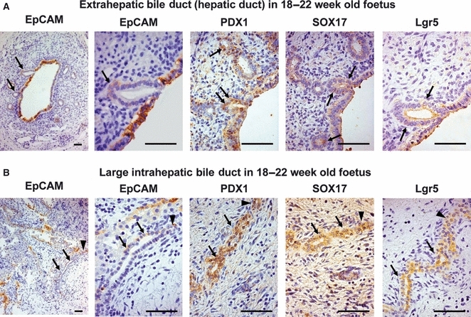
Stem/progenitors have been identified intrahepatically in the canals of Hering and extrahepatically in glands of the biliary tree. Glands of the biliary tree (peribiliary glands) are tubulo-alveolar glands with mucinous and serous acini, located deep within intrahepatic and extrahepatic bile ducts. We have shown that biliary tree stem/progenitors (BTSCs) are multipotent, giving rise in vitro and in vivo to hepatocytes, cholangiocytes or pancreatic islets. Cells with the phenotype of BTSCs are located at the bottom of the peribiliary glands near the fibromuscular layer. They are phenotypically heterogeneous, expressing transcription factors as well as surface and cytoplasmic markers for stem/progenitors of liver (e.g. SOX9/17), pancreas (e.g. PDX1) and endoderm (e.g. SOX17, EpCAM, NCAM, CXCR4, Lgr5, OCT4) but not for mature markers (e.g. albumin, secretin receptor or insulin). Subpopulations co-expressing liver and pancreatic markers (e.g. PDX1(+)/SOX17(+)) are EpCAM(+/-), and are assumed to be the most primitive of the BTSC subpopulations. Their descendants undergo a maturational lineage process from the interior to the surface of ducts and vary in the mature cells generated: pancreatic cells in hepatopancreatic ducts, liver cells in large intrahepatic bile ducts, and bile duct cells along most of the biliary tree. We hypothesize that there is ongoing organogenesis throughout life, with BTSCs giving rise to hepatic stem cells in the canals of Hering and to committed progenitors within the pancreas. The BTSCs are likely to be central to normal tissue turnover and injury repair and to be key elements in the pathophysiology of liver, pancreas and biliary tree diseases, including oncogenesis.


Figures

Fig. 1
Fig. 1
(A) Fetal common hepatic duct 18–22 weeks old (serial sections). The epithelia are composed of hepatopancreatic progenitors (EpCAM+/−, PDX1+, SOX17+, Lgr5+). Small evaginations of the epithelium, representing the bud of (peri)biliary glands, were observed (arrows). The PBG buds are composed of endodermal progenitors (arrows), mostly located at the bottom of the glands (see the arrow in the magnified image of cells stained with EpCAM). (B) In 18–22-week-old fetuses, the large intrahepatic bile duct can be found at the hilum. These ducts are composed mostly of endodermal (EpCAM+/−/PDX1+/SOX17+/Lgr5+) stem/progenitor cells (arrows). Scale bar: 50 μm.
Fig. 2
Fig. 2
Phenotype of glands within extrahepatic bile ducts. (A) In haematoxylin-eosin stains, (peri)biliary glands are tubulo-alveolar glands located in the lamina propria of duct walls (arrows). Immunohistochemistry showed the presence of cytokeratin-19 (CK 19)+ cells in the surface epithelium (cholangiocytes) and in the PBGs (arrows). (B) Immunohistochemistry for nuclear transcription factors and surface markers characterizing endodermal stem cells. Upper panels: cells within PBGs express markers typical of endodermal stem cells, such as FOXa2, CXCR4 and OCT4 (arrows). Lower panels: PBGs are composed of cells expressing typical markers of intestinal stem cell (Lgr5) and hepatic stem cell, such as EpCAM, NCAM and SOX9. Scale bar: 50 μm.
Fig. 3
Fig. 3
Triple/double immunofluorescence staining demonstrating the presence of endoderm-like and progenitor-like cells (BTSCs) and cells with an intermediate phenotype within PBGs. (A) EpCAM+/−/PDX1+/SOX17+ (or FOXa2+) cells were present in PBGs (arrows), representing ≍ 10% of the PBG cells. (B) Double immunofluorescence staining indicated the presence of a population of EpCAM+ cells which co-express markers of mature cells such as secretin receptor (cholangiocyte lineage), insulin (β-pancreatic cells) and, rarely, albumin (hepatocyte lineage). This intermediate compartment represents ≍ 20% of cells of PBGs at the hepatopancreatic ampulla. Scale bar: 50 μm.
Fig. 4
Fig. 4
Demonstration of the distinction in mature marker fate as one progresses along the biliary tree. (A,B) Double immunofluorescence for PDX1 and SOX19 and immunohistochemistry for cytokeratin-19, HepPar-1 and insulin at the level of the hepatopancreatic ampulla (A) and common hepatic/large intrahepatic bile ducts (B). Scale bar: 50 μm. Several differences could be noted. The presence of pancreatic progenitor-like cells (i.e. PDX1+/SOX17 cells, green arrows) and insulin+ cells was higher near the hepatopancreatic ampulla when compared with hepatic/large intrahepatic bile ducts. In contrast, there were more HepPar+ cells in common hepatic/large intrahepatic bile ducts than in the hepatopancreatic ampulla (see also Table 2).
Fig. 5
Fig. 5
Gradient in the expression of stem cell markers and of the proliferation of PBGs cells. (A–C) Immunohistochemistry for EpCAM counterstained with PAS. Glandular element just beneath the surface epithelium (see magnification in B) are mostly EpCAM and PAS+ (goblet cells); in contrast, acini located deep near the fibromuscular layer (see magnification in C) are composed of cells that are mostly EpCAM+ and PAS (arrows) or by EpCAM+/PAS+ cells (arrowhead). (D–F) Immunohistochemistry for PDX1. PDX1+ cells are mostly situated deep within duct walls (see magnification in F: arrows). PBGs near the surface epithelium are occasionally PDX1+ (arrow in E). (G–I) Immunohistochemistry for PCNA. Proliferating cells are mostly present in glandular elements located near the fibromuscular layer (I: arrows). Few cells are positive in more superficial acini. Notably, surface epithelial cells are mostly negative for PCNA. Scale bar: 50 μm.
Fig. 6
Fig. 6
Morphological aspects of glands within intrahepatic bile ducts. (A–B) Haematoxylin-eosin and PAS stains. PBGs were predominantly found at the level of large bile ducts; in these ducts, two types of (peri)biliary glands could be observed: intramural PBGs (arrows) were positioned inside the duct wall and appeared as shallow evaginations of the bile duct epithelium; extramural PBGs (box) were located outside the wall and were composed of PAS-negative cells. (C–D) Immunohistochemistry for CK19 and EpCAM showed the presence of positive cells in the surface epithelium, intra- (arrows) and extramural (box) PBGs. Scale bar: 50 μm.
Fig. 7
Fig. 7
Immunohistochemistry for PDX1, SOX17 and Lgr5 in intrahepatic bile ducts. (A) In large intrahepatic bile ducts, the surface epithelium and the intramural PBGs contain endoderm-like and progenitor-like cells (PDX1+/SOX17+/Lgr5+ cells), as indicated in seriated sections (arrows). (B) Extramural (peri)biliary glands are composed mostly of endoderm-like and progenitor-like cells, which co-express specific markers (PDX1+/SOX17+/Lgr5+). Notably, PDX1+/SOX19 cells were rarely found. Scale bar: 50 μm.

Similar articles

Cited by

References

    1. Alison MR, Golding MH, Sarraf CE. Pluripotential liver stem cells: facultative stem cells located in the biliary tree. Cell Prolif. 1996;29:373–402. - PubMed
    1. Antoniou A, Raynaud P, Cordi S, et al. Intrahepatic bile ducts develop according to a new mode of tubulogenesis regulated by the transcription factor SOX9. Gastroenterology. 2009;136:2325–2333. - PMC - PubMed
    1. Blechacz B, Komuta M, Roskams T, et al. Clinical diagnosis and staging of cholangiocarcinoma. Nat Rev Gastroenterol Hepatol. 2011;8:512–522. - PMC - PubMed
    1. Cardinale V, Semeraro R, Torrice A, et al. Intra-hepatic and extra-hepatic cholangiocarcinoma: new insight into epidemiology and risk factors. World J Gastrointest Oncol. 2010;2:407–416. - PMC - PubMed
    1. Cardinale V, Wang Y, Carpino G, et al. Multipotent stem/progenitor cells in human biliary tree give rise to hepatocytes, cholangiocytes and pancreatic islets. Hepatology. (in press) in press. - PubMed

Publication types