Comparative Study
. 2011 Dec 27;50(51):11058-69.
doi: 10.1021/bi201248b. Epub 2011 Dec 2.

Analysis of substrate access to active sites in bacterial multicomponent monooxygenase hydroxylases: X-ray crystal structure of xenon-pressurized phenol hydroxylase from Pseudomonas sp. OX1

Affiliations
Comparative Study

Analysis of substrate access to active sites in bacterial multicomponent monooxygenase hydroxylases: X-ray crystal structure of xenon-pressurized phenol hydroxylase from Pseudomonas sp. OX1

Michael S McCormick et al. Biochemistry. .

Erratum in

  • Biochemistry. 2012 Jan 10;51(1):573

Abstract

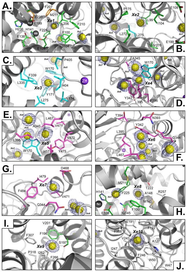
In all structurally characterized bacterial multicomponent monooxygenase (BMM) hydroxylase proteins, a series of hydrophobic cavities in the α-subunit trace a conserved path from the protein exterior to the carboxylate-bridged diiron active site. This study examines these cavities as a potential route for transport of dioxygen to the active site by crystallographic characterization of a xenon-pressurized sample of the hydroxylase component of phenol hydroxylase from Pseudomonas sp. OX1. Computational analyses of the hydrophobic cavities in the hydroxylase α-subunits of phenol hydroxylase (PHH), soluble methane monooxygenase (MMOH), and toluene/o-xylene monooxygenase (ToMOH) are also presented. The results, together with previous findings from crystallographic studies of xenon-pressurized sMMO hydroxylase, clearly identify the propensity for these cavities to bind hydrophobic gas molecules in the protein interior. This proposed functional role is supported by recent stopped flow kinetic studies of ToMOH variants [Song, W. J., et al. (2011) Proc. Natl. Acad. Sci. U.S.A.108, 14795-14800]. In addition to information about the Xe sites, the structure determination revealed significantly weakened binding of regulatory protein to the hydroxylase in comparison to that in the previously reported structure of PHH, as well as the presence of a newly identified metal-binding site in the α-subunit that adopts a linear coordination environment consistent with Cu(I), and a glycerol molecule bound to Fe1 in a fashion that is unique among hydrocarbon-diiron site adducts reported to date in BMM hydroxylase structures. Finally, a comparative analysis of the α-subunit structures of PHH, MMOH, and ToMOH details proposed routes for the other three BMM substrates, the hydrocarbon, electrons, and protons, comprising cavities, channels, hydrogen-bonding networks, and pores in the structures of their α-subunits.


Figures

Figure 1
Figure 1
Xenon binding in the α-subuint of Xe-PHH. Proposed substrate access pathways that are common to all structurally characterized BMMs are labeled with arrows. Van der Waals surfaces of the hydrophobic cavities and pore region are represented in magenta (cavity 3), cyan (cavity 2), green (cavity 1), and orange (pore region). The putative electron-transport hydrogen-bonding network residues are shown as yellow sticks. Iron and xenon atoms are represented as dark grey spheres and yellow spheres, respectively. The α-subunit protein backbone is shown as ribbons in light gray.
Figure 2
Figure 2
Detailed views of xenon binding sites in the α-subunit of Xe-PHH. Xenon (yellow), iron (dark gray), and zinc (purple) atoms are shown as spheres. Residues within 5 Å of xenon atoms are shown as sticks and colored according to the scheme used in Figure 1 if they contribute to van der Waals surface of the hydrophobic cavities or pore region, and in light grey if they do not. Composite omit electron density contoured to 5.0 sigma is shown in dark blue. The protein backbone is depicted as light grey ribbons. Xenon sites 1 through 10 are depicted (A through J).
Figure 3
Figure 3
Surface-to-active site pockets in the α-subuint of PHH. Hydrophobic cavities and pore region pockets are represented as van der Waals surfaces in magenta (cavity 3), cyan (cavity 2), green (cavity 1), and orange (pore region). Protein residues that contribute to the van der Waals surface are shown as sticks using the same color scheme as the surface representations. The putative electron-transport hydrogen-bonding network residues are shown as yellow sticks. Iron atoms are represented as dark grey spheres. The inset indicates the orientation of the hydroxylase dimer protomers A (dark ribbons) and B (light ribbons) depicted in the primary figure.
Figure 4
Figure 4
Surface-to-active site pockets in the α-subuint of MMOH. Hydrophobic cavities and pore region pockets are represented as van der Waals surfaces in magenta (cavity 3), cyan (cavity 2), green (cavity 1), and orange (pore region). Protein residues that contribute to the van der Waals surface are shown as sticks using the same color scheme as the surface representations. The putative electron-transport hydrogen-bonding network residues are shown as yellow sticks. Iron atoms are represented as dark grey spheres. The inset indicates the orientation of the hydroxylase dimer protomers A (dark ribbons) and B (light ribbons) depicted in the primary figure.
Figure 5
Figure 5
Surface-to-active site pockets in the α-subuint of ToMOH. Hydrophobic cavities, pore region, and channel pockets are represented as van der Waals surfaces in magenta (cavity 3), cyan (cavity 2), green (cavity 1), orange (pore region), and grey (channel). Protein residues that contribute to the van der Waals surface are shown as sticks using the same color scheme as the surface representations. The putative electron-transport hydrogen-bonding network residues are shown as yellow sticks. Iron atoms are represented as dark grey spheres. The inset indicates the orientation of the hydroxylase dimer protomers A (dark ribbons) and B (light ribbons) depicted in the primary figure.
Figure 6
Figure 6
View down the pore pathway into the active site cavity in Xe-PHH (A), MMOH (B), and ToMOH (C). Hydrophobic cavities pockets and the ToMOH channel are represented as van der Waals surfaces in magenta (cavity 3), cyan (cavity 2), green (cavity 1), and grey (channel). Protein residue side chains that comprise the pore pathway are shown as sticks in orange (carbon), blue (nitrogen), and red (oxygen). The four-helix bundle is represented as ribbons in light grey, and iron ligands (green) and the surface-to diiron center hydrogen bonding network residues (yellow) are shown as lines.
Figure 7
Figure 7
Diiron center access routes in the α-subunit of PHH (A), PHH-PHM (B), MMOH (C), and ToMOH (D) as calculated by CAVER. Substrate pathways are represented as surfaces in magenta (hydrophobic cavities), orange (pore region), and gray (channel). Hydroxylase α-subunit is shown as light grey ribbons; PHM is shown as blue ribbons (B only). Iron and zinc (A and B only) atoms are shown as dark gray and purple spheres, respectively.

Similar articles

Cited by

References

    1. Cafaro V, Izzo V, Scognamiglio R, Notomista E, Capasso P, Casbarra A, Pucci P, Di Donato A. Phenol Hydroxylase and Toluene/o-Xylene Monooxygenase from Pseudomonas stutzeri OX1: Interplay Between Two Enzymes. Appl Environ Microbiol. 2004;70:2211–2219. - PMC - PubMed
    1. Sazinsky MH, Lippard SJ. Correlating structure with function in bacterial multicomponent monooxygenases and related diiron proteins. Acc Chem Res. 2006;39:558–566. - PubMed
    1. Leahy JG, Batchelor PJ, Morcomb SM. Evolution of the soluble diiron monooxygenases. FEMS Microbiol Rev. 2003;27:449–479. - PubMed
    1. Rosenzweig AC, Brandstetter H, Whittington DA, Nordlund P, Lippard SJ, Frederick CA. Crystal Structures of the Methane Monooxygenase Hydroxylase from Methylococcus capsulatus (Bath): Implications for Substrate Gating and Component Interactions. Proteins. 1997;29:141–152. - PubMed
    1. Rosenzweig AC, Frederick CA, Lippard SJ, Nordlund P. Crystal structure of a bacterial non-hame iron hydroxylase that catalyzes the biological oxidation of methane. Nature. 1993;366:537–543. - PubMed

Publication types

MeSH terms

Associated data