. 2012 Jan 1;9(1):187-95.
doi: 10.1021/mp200461s. Epub 2011 Dec 12.

Improved coiled-coil design enhances interaction with Bcr-Abl and induces apoptosis

Affiliations

Improved coiled-coil design enhances interaction with Bcr-Abl and induces apoptosis

Andrew S Dixon et al. Mol Pharm. .

Erratum in

Abstract

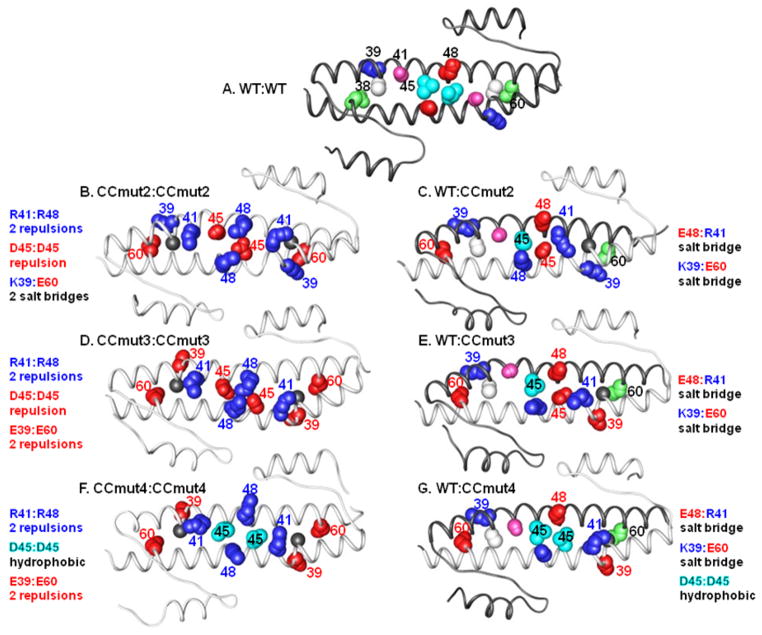
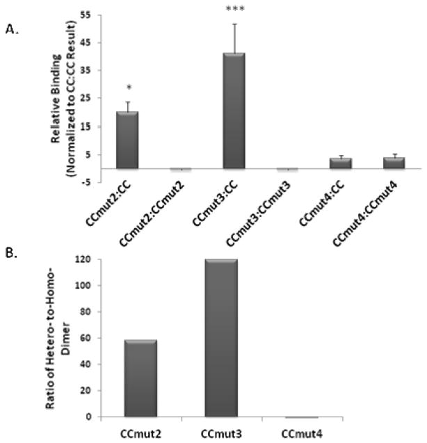
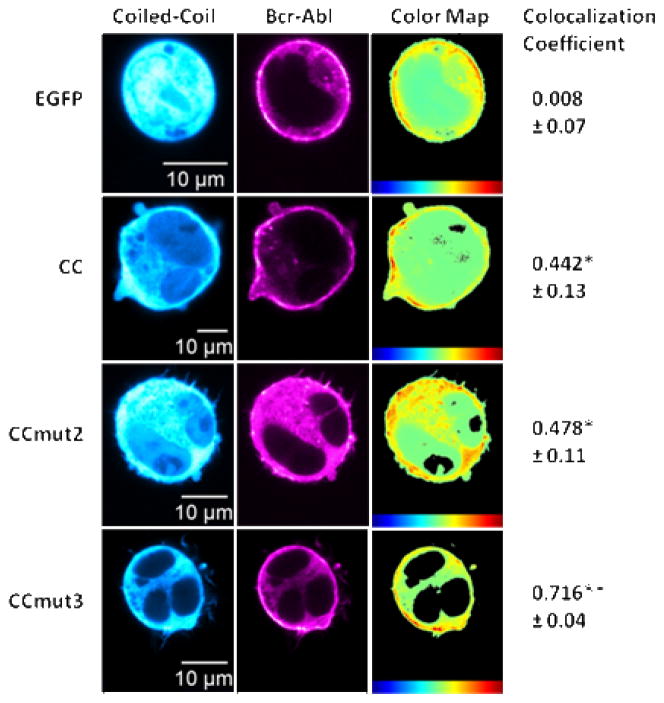
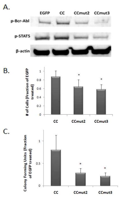
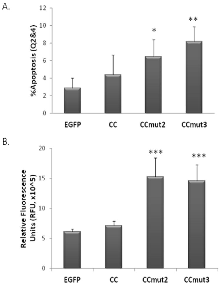
The oncoprotein Bcr-Abl drives aberrant downstream activity through trans-autophosphorylation of homo-oligomers in chronic myelogenous leukemia (CML).(1, 2) The formation of Bcr-Abl oligomers is achieved through the coiled-coil domain at the N-terminus of Bcr.(3, 4) We have previously reported a modified version of this coiled-coil domain, CCmut2, which exhibits disruption of Bcr-Abl oligomeric complexes and results in decreased proliferation of CML cells and induction of apoptosis.(5) A major contributing factor to these enhanced capabilities is the destabilization of the CCmut2 homodimers, increasing the availability to interact with and inhibit Bcr-Abl. Here, we included an additional mutation (K39E) that could in turn further destabilize the mutant homodimer. Incorporation of this modification into CCmut2 (C38A, S41R, L45D, E48R, Q60E) generated what we termed CCmut3, and resulted in further improvements in the binding properties with the wild-type coiled-coil domain representative of Bcr-Abl [corrected]. A separate construct containing one revert mutation, CCmut4, did not demonstrate improved oligomeric properties and indicated the importance of the L45D mutation. CCmut3 demonstrated improved oligomerization via a two-hybrid assay as well as through colocalization studies, in addition to showing similar biologic activity as CCmut2. The improved binding between CCmut3 and the Bcr-Abl coiled-coil may be used to redirect Bcr-Abl to alternative subcellular locations with interesting therapeutic implications.


Figures

Figure 1
Figure 1
Ribbon diagrams of the coiled-coil domains. White ribbons indicate wild type (WT) coiled-coil, grey ribbons indicate mutant coiled-coils, and each homo-dimer (A, B, D, and F) or hetero-dimer (C, E, and G) is labeled above. Blue numbering/spheres indicates positively charged amino acid residue; red numbering/spheres indicates negatively charged amino acid residue. For the WT, white = C38, blue (+ chg) = K39, purple = S41, cyan (hφ) = L45, red (− chg) = E48, green = Q60. Underlined residue is colored in the figure. For CC mutants, gray = C38A, red (− chg) = K39E, blue (+ chg) = S41R, red (− chg) = L45D, blue (+ chg) = E48R, red (− chg) = Q60E. Underlined residue is colored in the figure. A) WT:WT homo-dimer. Only the top strand is numbered. B) CCmut2:CCmut2 homo-dimer (CCmut2 contains C38A, S41R, L45D, E48R, Q60E mutations). The two R41:R48, and one D45:D45 charge-charge repulsion are shown, as well as the two sets of K39:E60 salt bridges. C) WT:CCmut2 hetero-dimer. The E48:R41 and K39:E60 salt bridges are indicated. D) CCmut3:CCmut3 homo-dimer (CCmut3 contains C38A, K39E, S41R, L45D, E48R, Q60E mutations). The two sets of K39:E60 salt bridges are now replaced with two sets of E39:E60 charge-charge repulsions. The two R41:R48 and one D45:D45 charge-charge repulsions are retained. E) WT:CCmut3 hetero-dimers. CCmut3 may form E48:R41 and K39:E60 salt bridges with WT as illustrated. F) CCmut4 homo-dimers (CCmut4 contains C38A, K39E, S41R, E48R, Q60E mutations). Similar to CCmut3 homo-dimer, the two sets of E39:E60, and two R41:R48 charge-charge repulsions are again illustrated. D45:D45 charge-charge repulsion is now replaced with a L45:L45 hydrophobic interaction in the middle. G) WT:CCmut4 hetero-dimers. The K39:E60 and E48:R41 salt bridges, and L45:L45 hydrophobic interactions are shown.
Figure 2
Figure 2
Mammalian two-hybrid assay. A) Relative response ratios determined for each interaction and normalized to the wild-type homo-oligomerization (CC:CC) to indicate the relative binding efficiency. Values indicated are means ± S.D. Statistics were performed on the values prior to normalizing to CC so as to include the wild-type interaction in the statistics (n=4 or 5). *p<0.05, ***p<0.01 versus CC:CC interaction; one-way ANOVA, Tukey’s post test. B) The quotient of the hetero-dimer and homo-dimer inte raction (absolute value) is graphed for all three mutants, indicating the preferential binding to CC over the formation of a homo-dimer.
Figure 3
Figure 3
Colocalization with Bcr-Abl. Representative images of either EGFP or coiled-coil domain seen in left column (false colored cyan for visualization) with representative images of Bcr-Abl distribution in middle column (false colored magenta for visualization). Heat maps indicating the colocalization of the two fluorophores seen in the right column with colocalization scale at the bottom (red highest) followed by the colocalization coefficient. The mean colocalization coefficient was determined from at least three cells per transfection (n=4), with values reported as means ± S.E.M. *p<0.05, **p<0.01 control; one-way ANOVA, Tukey’s post test.
Figure 4
Figure 4
Effects of CCs on Bcr-Abl and proliferation of K562 cells. A) Western blot indicating the phosphorylation of Bcr-Abl (top, p-Bcr-Abl) and STAT5 (middle, p-STAT5). B) Colony forming units counted seven days after seeding 103 transfected K562 cells into methylcellulose medium, normalized to colony growth from EGFP control (n=3). Values reported as means ± S.D. *p<0.05 control; one-way ANOVA, Tukey’s post test. C) Viable K562 cells were counted 48 hrs following transfection, with the number of proliferating cells normalized to the number resulting from the EGFP transfection (n=4). Values reported as means ± S.D. *p<0.05 control; one-way ANOVA, Tukey’s post test.
Figure 5
Figure 5
Apoptosis assays. A) 48 hrs following transfection into K562 cells phosphatidylserine externalization was assessed through flow cytometry. After gating on EGFP fluorescence to select only the transfected cells, the number of 7AAD/Annexin+ cells (Q2, early apoptosis) was combined with the 7AAD+/Annexin+ cells (Q4, late apoptosis) and used to determine the percentage of apoptotic cells (n=3). Values reported as means ± S.D. *p<0.05, **p<0.01 control; one-way ANOVA, Tukey’s post test. B) 48 hrs following transfection into K562 cells the activation of caspase-3/7 was analyzed in the cell lysates through a fluorescence-based assay (n=3). Values reported as means ± S.D. ***p<0.01; one-way ANOVA, Tukey’s post test.
Figure 6
Figure 6
Nuclear segmentation of K562 cells. A) Representative images of K562 cells 48 hrs after transfection of EGFP (top row) or CCmut3 (bottom row). Left column = phase contrast images with black arrow indicating example of cell shrinkage; middle column = fluorescence from EGFP; right column = stained nuclei with white arrow indicating example of nuclear segmentation. B) Quantitative results of nuclear segmentation. Percentage of transfected cells with segmented nuclei was determined from three or four fields of view, and repeated with three separate transfections (n=3). Values reported as means ± S.D. *p<0.05; one-way ANOVA, Tukey’s post test.

Similar articles

Cited by

References

    1. McWhirter JR, Galasso DL, Wang JY. A coiled-coil oligomerization domain of Bcr is essential for the transforming function of Bcr-Abl oncoproteins. Mol Cell Biol. 1993;13(12):7587–95. - PMC - PubMed
    1. Guo XY, Cuillerot JM, Wang T, Wu Y, Arlinghaus R, Claxton D, Bachier C, Greenberger J, Colombowala I, Deisseroth AB. Peptide containing the BCR oligomerization domain (AA 1-160) reverses the transformed phenotype of p210bcr-abl positive 32D myeloid leukemia cells. Oncogene. 1998;17(7):825–33. - PubMed
    1. Taylor CM, Keating AE. Orientation and oligomerization specificity of the Bcr coiled-coil oligomerization domain. Biochemisty. 2005;44(49):16246–56. - PMC - PubMed
    1. Zhao X, Ghaffari S, Lodish H, Malashkevich VN, Kim PS. Structure of the Bcr-Abl oncoprotein oligomerization domain. Nat Struct Biol. 2002;9(2):117–20. - PubMed
    1. Dixon AS, Pendley SS, Bruno BJ, Woessner DW, Shimpi AA, Cheatham TE, 3rd, Lim CS. Disruption of BCR-ABL coiled coil oligomerization by design. J Biol Chem. 2011;286(31):27751–60. - PMC - PubMed

Publication types

MeSH terms