. 2011 Dec 2;9(6):541-52.
doi: 10.1016/j.stem.2011.10.003.

Erythroid/myeloid progenitors and hematopoietic stem cells originate from distinct populations of endothelial cells

Affiliations

Erythroid/myeloid progenitors and hematopoietic stem cells originate from distinct populations of endothelial cells

Michael J Chen et al. Cell Stem Cell. .

Erratum in

  • Cell Stem Cell. 2012 Mar 2;10(3):345-6

Abstract

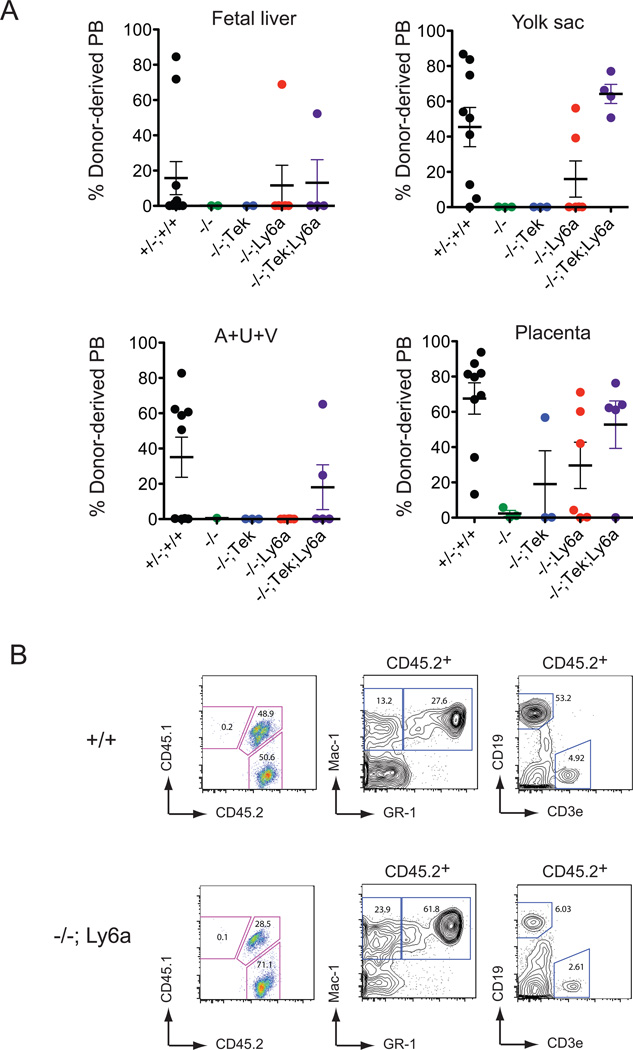
Hematopoietic stem cells (HSCs) and an earlier wave of definitive erythroid/myeloid progenitors (EMPs) differentiate from hemogenic endothelial cells in the conceptus. EMPs can be generated in vitro from embryonic or induced pluripotent stem cells, but efforts to produce HSCs have largely failed. The formation of both EMPs and HSCs requires the transcription factor Runx1 and its non-DNA binding partner core binding factor β (CBFβ). Here we show that the requirements for CBFβ in EMP and HSC formation in the conceptus are temporally and spatially distinct. Panendothelial expression of CBFβ in Tek-expressing cells was sufficient for EMP formation, but was not adequate for HSC formation. Expression of CBFβ in Ly6a-expressing cells, on the other hand, was sufficient for HSC, but not EMP, formation. The data indicate that EMPs and HSCs differentiate from distinct populations of hemogenic endothelial cells, with Ly6a expression specifically marking the HSC-generating hemogenic endothelium.


Figures

Figure 1
Figure 1. Expression of GFP/CBFβ from the Tek regulatory sequences poorly rescues lymphoid progenitor and HSC formation
A. Fetal liver progenitors (LinKit+Flt3+) were sorted from 14.5 dpc embryos and plated on OP9 or OP9-DL4 stromal cells. Number of cells plated were 400 and 200 Tek-GFP/Cbfb on OP9 and OP9-DL1, respectively; 2000 Cbfb−/−; Tek-GFP/Cbfb on both OP9 and OP9-DL4. OP9 cultures contained 5 ng/ml Flt3L, 10ng/ml IL7. OP9-DL4 cultures contained 5 ng/ml Flt3L, 1 ng/ml IL7. Cells were analyzed for the expression of lymphoid markers 7 days later. B. Limit dilution analysis of fetal liver progenitors cultured as described above. C. Percent and number of recipients containing ≥ 5% donor-derived blood 16 weeks following transplantation with 2 × 10⁵, 5 × 10⁵, or 1 × 10 6 14.5 dpc fetal liver cells from wild type (+/+), Cbfb−/− (−/−), and Cbfb−/−; Tek-GFP/Cbfb (−/−;Tek) conceptuses. D. Representative scatter plots of 13.5 dpc fetal liver HSCs (LSK cells). E. Total number of fetal liver and LSK cells at 13.5 dpc. Data are compiled from 2–5 fetuses. Error bars represent standard error of the mean (SEM). F. Representative histograms showing GFP/CBFβ expression in the LSK and LT-HSC populations of 13.5 dpc Tek-GFP/Cbfb fetal livers. The two green traces and percentages in the histograms represent two independent fetuses.
Figure 2
Figure 2. Expression of GFP/CBFβ in endothelial cells and hematopoietic cells from the Ly6a-GFP/Cbfb transgene
A. Western blot analysis of whole bone marrow from wild type, Ly6a-GFP, and three independent Ly6a-GFP/Cbfb lines using a monoclonal antibody recognizing CBFβ. Endogenous CBFβ and the GFP/CBFβ fusion protein are indicated. Whole cell lysates from 2 × 10⁵ cells were loaded. B. Representative histograms showing GFP/CBFβ expression in the LSK and LT-HSC populations in the bone marrow (BM) of thee independent lines of adult Ly6a-GFP/Cbfb mice. C. (Left) Whole mount immunofluorescence of an 11.5 dpc Ly6a-GFP/Cbfb conceptus. The fetus is outlined with a dashed white line. Pl, placenta; UA, umbilical artery. (Right) An isolated AGM region with expression in the dorsal aorta (DA), vitelline artery (VA), UA, mesonephric tubules (MT), and Wolffian/Mullerian ducts (W/M), identical to that reported for the Ly6a-GFP transgene (de Bruijn et al., 2002). D. Representative histograms showing GFP/CBFβ expression in the majority phenotypic LT-HSC in a 13.5 dpc Ly6a-GFP/Cbfb fetal liver (FL, Line A).
Figure 3
Figure 3. EMPs in Cbfb−/− conceptuses are rescued upon expression of GFP/CBFβ from the Tek but not the Ly6a-driven transgene. See also Figure S1
Shown are the numbers of hematopoietic progenitors (colonies) per embryo equivalent (ee) in four hematopoietic tissues: fetal liver, yolk sac, AGM region combined with umbilical and vitelline arteries (A+U+V), and placenta. Numbers are compiled from 3–14 11.5 or 14.5 dpc conceptuses per genotype. P values in boxes indicate significant differences in total progenitor numbers in Cbfb−/− conceptuses (−/−, indicated by #) versus Cbfb−/− conceptuses carrying transgenes as determined by one-way ANOVA. Error bars represent SEM. Asterisks indicate significant differences compared to −/− (P < 0.01) according to Dunnett’s Multiple Comparison test. Differences between Cbfb−/−, Cbfb−/−; Tek-GFP/Cbfb (−/−; Tek) and Cbfb−/−; Ly6a-GFP/Cbfb (−/−; Ly6a) were determined by unpaired two-tailed Student’s t test. Cbfb−/−; Tek-GFP/Cbfb; Ly6a-GFP/Cbfb is −/−; Tek+Ly6a.
Figure 4
Figure 4. Expression of GFP/CBFβ from the Ly6a regulatory sequences rescues HSCs in Cbfb−/− conceptuses
A. Repopulation by 0.9 ee of 11.5 dpc cells from conceptuses of the indicated genotypes at 4 months post transplant. Percent engraftment represents the percent donor-derived blood; each dot is an individual transplant recipient. Error bars represent SEM. B. Multi-lineage engraftment by donor derived wild type and Cbfb−/−; Ly6a-GFP/Cbfb HSCs in a recipient of placenta cells.
Figure 5
Figure 5. Expression of the Tek-GFP/Cbfb and Ly6a-GFP/Cbfb transgenes in 8.5–9.5 dpc conceptuses analyzed by confocal microscopy. See also Figure S2 and Movie S2
A. Whole mount immunofluorescence of a 7s Tek-GFP/Cbfb conceptus using antibodies recognizing CD31 and GFP (10X). Scale bars for all panels = 100µm. Figure represents the MAX of six consecutive 3µm z-sections for CD31 and GFP. DA, dorsal aorta; YS, yolk sac; Al, allantois; Pl, placenta; H, heart. B. A 11s Ly6a-GFP/Cbfb conceptus, analyzed as described in A. C. A 16s Tek-GFP/Cbfb conceptus reconstructed as the standard deviation (STD) of 13 consecutive 1.3µm z-sections for CD31, and the SUM for GFP (10X). D. A 16s Ly6a-GFP/Cbfb conceptus, analyzed as described in C. Expression is observed in endothelial cells in the caudal aspect of the dorsal aortae, placenta, yolk sac (primarily caudal region), vitelline artery (VA), vitelline vein (VV), foregut (Fg), and in the hindgut (not shown). See also Movie S1.
Figure 6
Figure 6. Expression of the Tek-GFP/Cbfb and Ly6a-GFP/Cbfb transgenes in 10.5 dpc conceptuses. See also Figures S3, S5
A. Whole mount immunofluorescence of a 36s Tek-GFP/Cbfb specimen using antibodies recognizing CD31 and GFP (5X). Scale bars = 100µm. View represents AVG of fourteen 10µm z-sections. DA, dorsal aorta; UA, umbilical artery; VA, vitelline artery, Fl, fetal liver. B. A 38s Ly6a-GFP/Cbfb specimen, analyzed as described in A. The Ly6a-GFP/Cbfb transgene is also highly expressed in the tail. MT, Mullerian tubules; HG, hindgut. See also Movie S3. C. Yolk sac from the Tek-GFP/Cbfb fetus shown in A (5X). View represents SUM of 32 9.7µm z-sections. Asterisk shows area of expression in the yolk sac vascular plexus. D. Yolk sac from the Ly6a-GFP/Cbfb fetus shown in B, analyzed as described in C. Arrows show expression in large vitelline vessels. E. Umbilical artery from the Tek-GFP/Cbfb fetus shown in A (20X). View represents SUM of six 0.38µm z-sections. F. Umbilical artery from the Ly6a-GFP/Cbfb fetus shown in B (20X), analyzed as described in E. G. Intra-arterial hematopoietic cluster from the umbilical artery of a Tek-GFP/Cbfb fetus (20X). H. Intra-arterial hematopoietic cluster from the umbilical artery of a Ly6a-GFP/Cbfb fetus, as in G.
Figure 7
Figure 7. Ly6a transgene expression marks a subset of CD144+, ESAM+, CD105+, Tek+, Flk1+, and c-Kit+ cells. See also Figure S4
A. Live (DAP1 negative) cells from the 10.5 dpc A+U+V were gated for GFP/CBFβ in the plot furthest to the left, and GFP/CBFβ+ cells are shown as red dots in this and all other plots. B. Live cells are gated for CD31 and c-Kit expression and divided into endothelial cells (CD31+ c-Kit) and hematopoietic cells (CD31+ c-Kit+). Each population in turn is analyzed for ESAM and CD144 expression, and all ESAM+ cells for CD144 and GFP/CBFβ expression. Black numbers represent the percentage of all cells in a particular gate, while red numbers represent percentages of GFP/CBFβ+ cells in that gate. C. Analysis of CD105+, Tek+, and Flk1+ populations, gated as described in A,B. D. Diagram illustrating that Ly6a-GFP expression marks the HSC-producing population of Runx1+ hemogenic endothelium.

References

    1. Adja N, Stacy T, Speck NA, Liu PP. The leukemic protein CBFβ-SMMHC sequesters CBFα2 into cytoskeletal filaments and aggregates. Mol Cell Biol. 1998;18:7432–7443. - PMC - PubMed
    1. Bertrand JY, Chi NC, Santoso B, Teng S, Stainier DY, Traver D. Haematopoietic stem cells derive directly from aortic endothelium during development. Nature. 464:108–111. - PMC - PubMed
    1. Boisset JC, van Cappellen W, Andrieu-Soler C, Galjart N, Dzierzak E, Robin C. In vivo imaging of haematopoietic cells emerging from the mouse aortic endothelium. Nature. 2010;464:116–120. - PubMed
    1. Bollerot K, Romero S, Dunon D, Jaffredo T. Core binding factor in the early avian embryo: cloning of Cbfbeta and combinatorial expression patterns with Runx1. Gene Expr Patterns. 2005;6:29–39. - PubMed
    1. Cai Z, de Bruijn MFTR, Ma X, Dortland B, Luteijn T, Downing JR, Dzierzak E. Haploinsufficiency of AML1/CBFA2 affects the embryonic generation of mouse hematopoietic stem cells. Immunity. 2000;13:423–431. - PubMed

Publication types

MeSH terms