

Gastric schwannoma: a clinicopathologic study of 51 cases and critical review of the literature
- PMID: 22137423
- PMCID: PMC3305846
- DOI: 10.1016/j.humpath.2011.07.006
Gastric schwannoma: a clinicopathologic study of 51 cases and critical review of the literature
Abstract
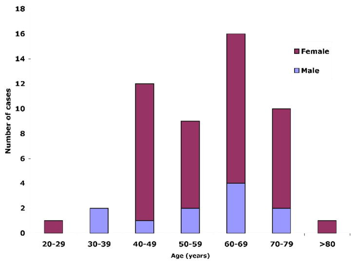
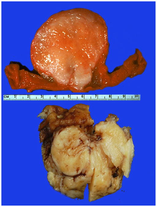
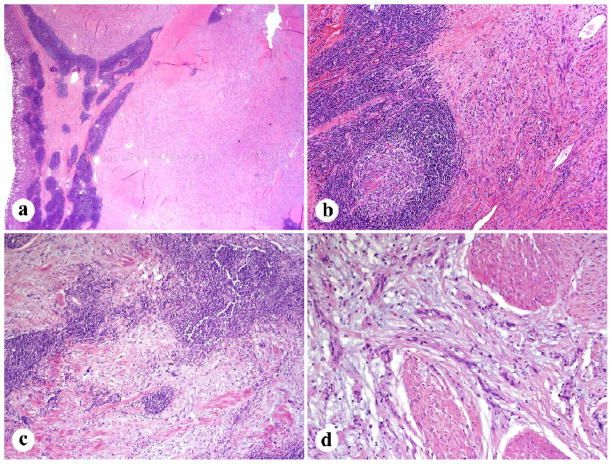
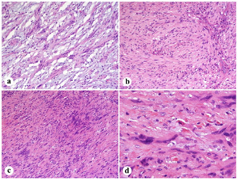
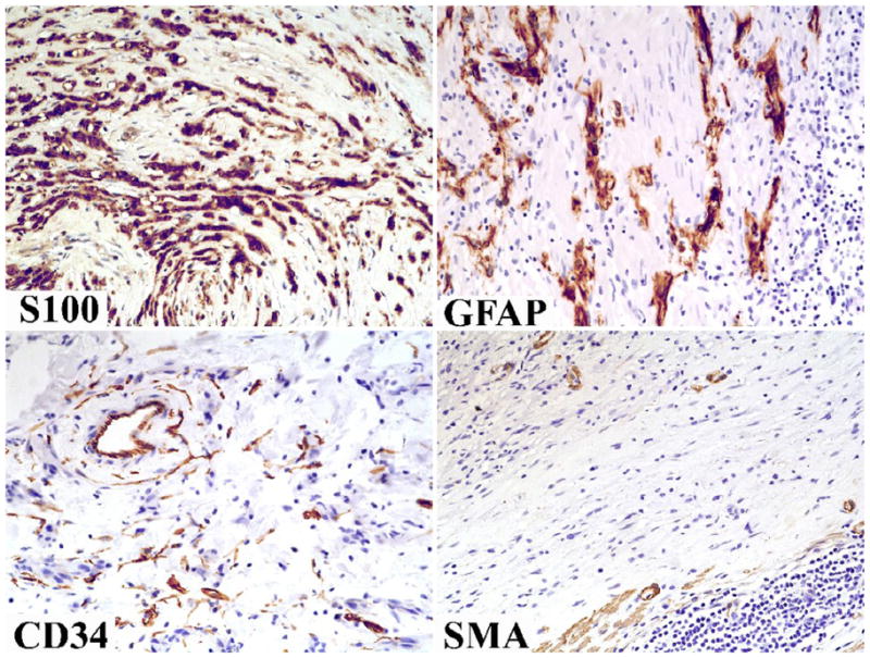
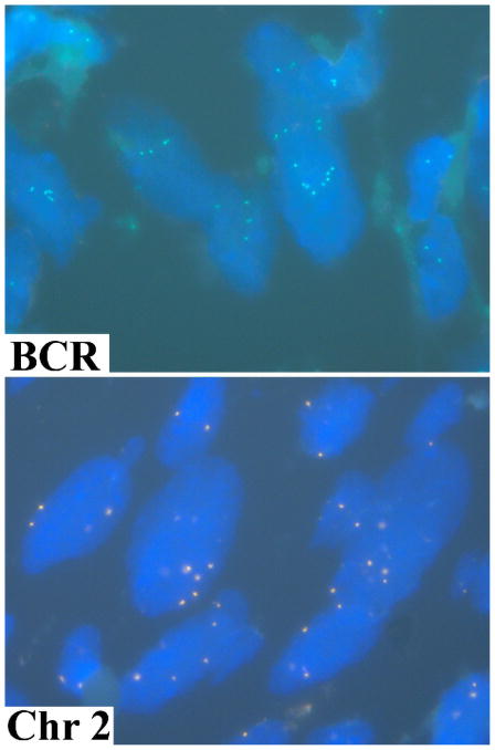
Schwannoma is a rare gastrointestinal mesenchymal tumor, as the vast majority of gastric mesenchymal tumors are gastrointestinal stromal tumors. In this study, we analyzed clinicopathologically 51 gastric schwannomas. These tumors predominantly occurred in older adults with a marked female predominance (40 women and 11 men; median and mean ages, 60 and 58 years). They variably presented with gastric discomfort, bleeding, or rarely gastric outlet obstruction; and many were incidental findings during other medical procedures. The tumors ranged from 1 to 10.5 cm (median, 4.5 cm). The typical histologic features included spindle cells usually with microtrabecular architecture and focal nuclear atypia, and peritumoral lymphoid cuff, whereas features of soft tissue schwannomas, such as encapsulation, nuclear palisading, vascular hyalinization, and dilatation, were absent or infrequent. Median mitotic count was 2/50 high-power fields, with the highest count being 13/50 high-power fields. No malignant variants were recognized, and long-term follow-up did not reveal recurrences or metastases. Immunohistochemically, all examined tumors were S100 protein positive and most were also GFAP positive, whereas CD34 and NF68 were encountered rarely and all tumors were negative for HMB45, KIT, DOG1/Ano 1, smooth muscle actin, desmin, and synaptophysin. None of the 9 tumors studied contained gastrointestinal stromal tumor-specific KIT or PDGFRA mutations. Fluorescence in situ hybridization studies revealed multiple signals with BCR probe (chromosome 22) and centromeric probes for chromosomes 2 and 18 suggesting polyploidy. These findings indicate that gastric schwannoma is a distinctive form of peripheral nerve sheath tumor that in many ways differs from soft tissue schwannoma. It should be distinguished from gastrointestinal stromal tumor and other mesenchymal tumors of the gastrointestinal tract, such as the S100 protein-positive gastrointestinal clear cell sarcoma and metastatic melanoma.
Published by Elsevier Inc.
Figures
References
-
- Daimaru Y, Kido H, Hashimoto H, et al. Benign schwannoma of the gastrointestinal tract: A clinicopathologic and immunohistochemical study. Hum Pathol. 1988;19:257–64. - PubMed
-
- Agaimy A, Markl B, Kitz J, et al. Peripheral nerve sheath tumors of the gastrointestinal tract: a multicenter study of 58 patients including NF1-associated gastric schwannoma and unusual morphological variants. Virchows Arch. 2010;456:411–22. - PubMed
-
- Hong HS, Ha HK, Won HJ, et al. Gastric schwannomas: radiologic features with endoscopic and pathologic correlation. Clinical Radiology. 2008;63:536–42. - PubMed
-
- Jung MK, Jeon SW, Cho CM, et al. Gastric schwannomas: endoscopic characteristics. Abdom Imaging. 2008;33:388–90. - PubMed
-
- Prevot S, Bienvenu L, Vaillant JC, et al. Benign schwannoma of the digestive tract. A clinicopathologic and immunohistochemical study of five cases, including a case of esophageal tumor. Am J Surg Pathol. 1999;23:431–6. - PubMed
Publication types
MeSH terms
Substances
Grants and funding
LinkOut - more resources
Full Text Sources
Medical
Miscellaneous
