. 2011 Dec 23;44(6):878-92.
doi: 10.1016/j.molcel.2011.11.005. Epub 2011 Dec 1.

Chemical genetic screen for AMPKα2 substrates uncovers a network of proteins involved in mitosis

Affiliations

Chemical genetic screen for AMPKα2 substrates uncovers a network of proteins involved in mitosis

Max R Banko et al. Mol Cell. .

Abstract

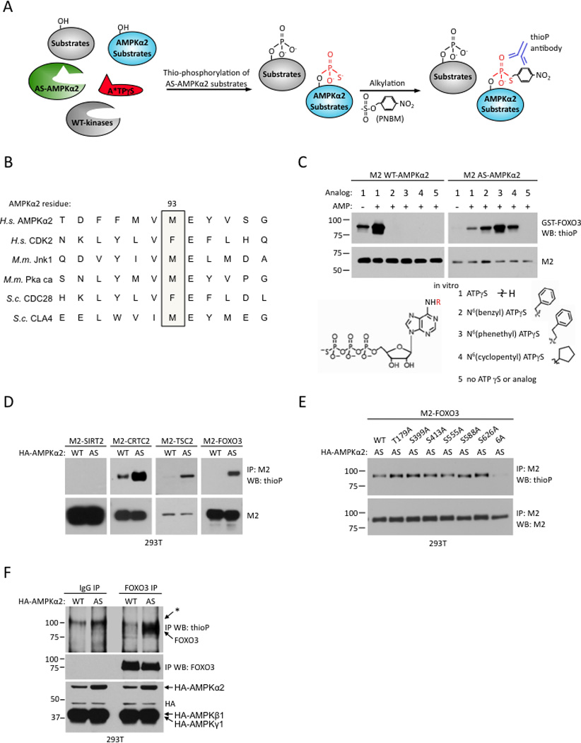
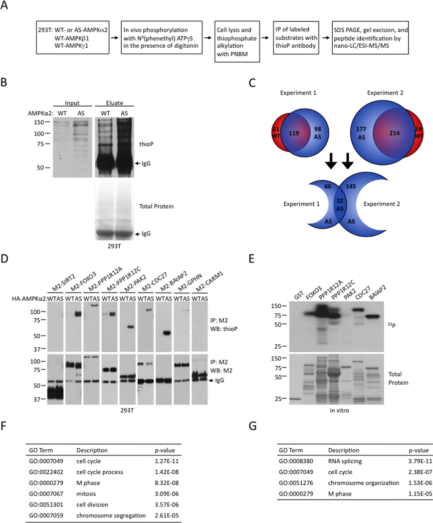
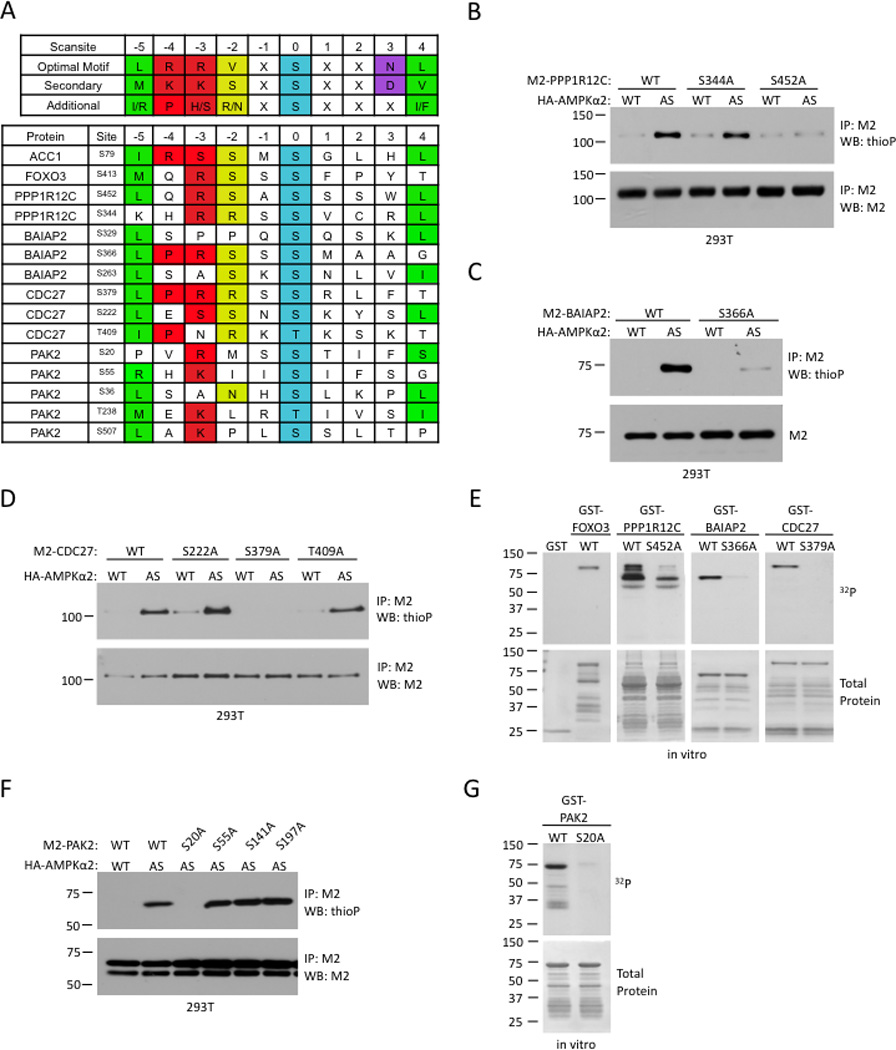
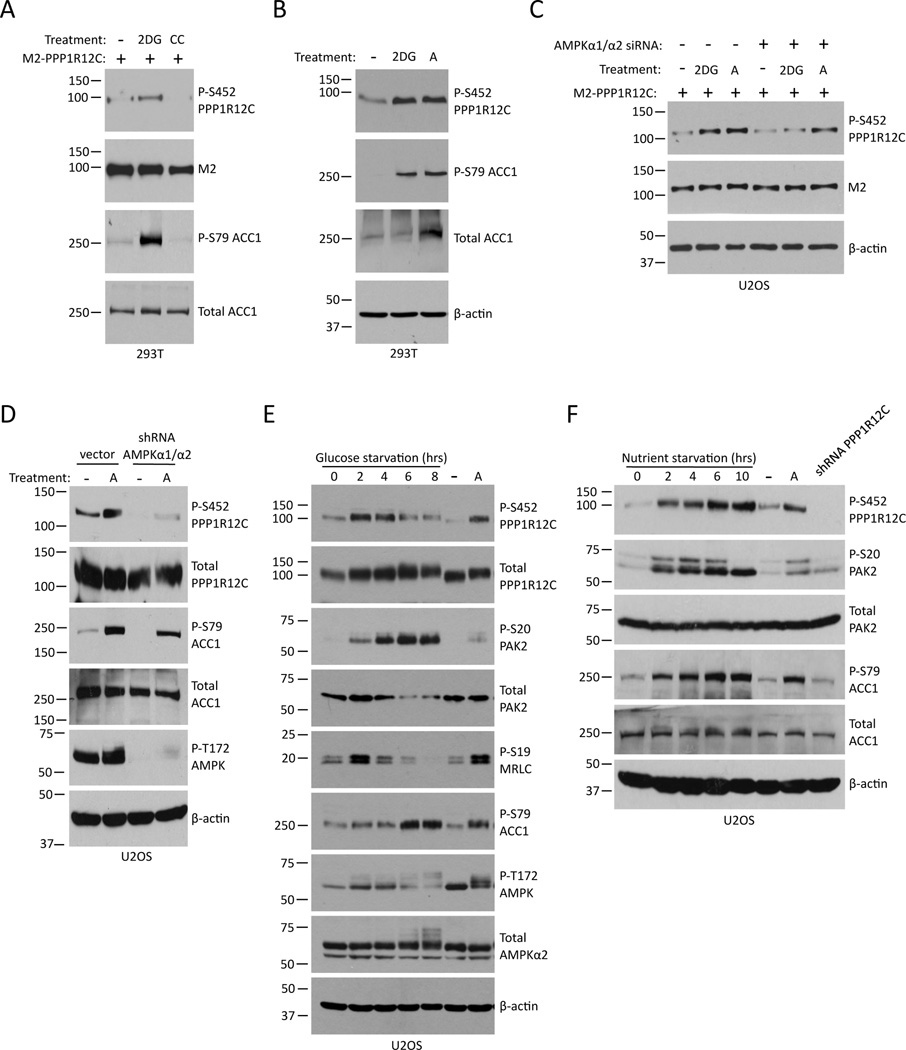
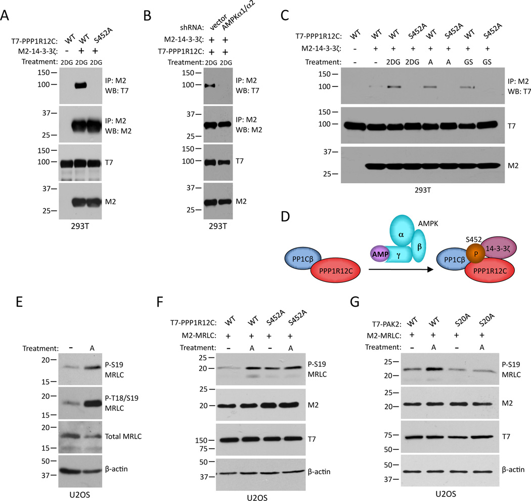
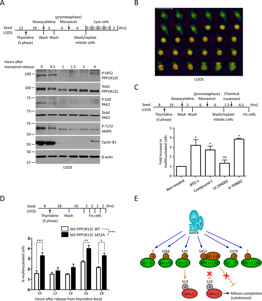
The energy-sensing AMP-activated protein kinase (AMPK) is activated by low nutrient levels. Functions of AMPK, other than its role in cellular metabolism, are just beginning to emerge. Here we use a chemical genetics screen to identify direct substrates of AMPK in human cells. We find that AMPK phosphorylates 28 previously unidentified substrates, several of which are involved in mitosis and cytokinesis. We identify the residues phosphorylated by AMPK in vivo in several substrates, including protein phosphatase 1 regulatory subunit 12C (PPP1R12C) and p21-activated protein kinase (PAK2). AMPK-induced phosphorylation is necessary for PPP1R12C interaction with 14-3-3 and phosphorylation of myosin regulatory light chain. Both AMPK activity and PPP1R12C phosphorylation are increased in mitotic cells and are important for mitosis completion. These findings suggest that AMPK coordinates nutrient status with mitosis completion, which may be critical for the organism's response to low nutrients during development, or in adult stem and cancer cells.


Figures

Figure 1
Figure 1. Generation of an analog-specific version of AMPKα2
(A) Strategy for labeling substrates of analog-specific AMPKα2 (AS-AMPKα2). A*TPγS: bulky ATP analog. PNBM: p-nitrobenzyl mesylate. (B) Identification of the gatekeeper amino acid in the ATP-binding pocket of human AMPKα2 by alignment with other kinases for which analog-specific mutants have been created (Allen et al., 2007; Niswender et al., 2002; Polson et al., 2001; Ubersax et al., 2003; Ventura et al., 2006; Weiss et al., 2000). H.s.: Homo sapiens; M.m.: Mus musculus; S.c: Saccharomyces cerevisiae. (C) In vitro kinase assay using WT- or AS-AMPKα2 immunoprecipitated from 293T cells with GST-FOXO3 as a substrate and the indicated N6-substituted ATPγS analog as a phosphodonor. Red R: site of N6-alkyl group attachment 1–4 indicated at right. Western blots representative of 2 independent experiments. (D) AS-AMPKα2 phosphorylates known AMPK substrates in cells in the presence of N6-(phenethyl) ATPγS. Substrates were immunoprecipitated with the antibodies to M2. Western blots representative of 2 independent experiments. (E) AS-AMPKα2 no longer phosphorylates M2-FOXO3 with the point mutations in the AMPK phosphorylation sites in cells. 6A: T179A/S399A/S413A/S55A/S588/S626A. (F) AS-AMPKα2 can phosphorylate endogenous FOXO3 in 293T cells. *: background band present in all samples. Western blots representative of 3 independent experiments.
Figure 2
Figure 2. Identification of direct substrates of AMPKα2 in cells by a chemical genetic screen
(A) Strategy for identifying endogenous direct substrates of AS-AMPKα2 in cells. (B) Phosphorylation of endogenous AMPKα2 substrates in 293T cells expressing WT- or AS-AMPKα2. Left panel: western blot of lysates run before immunoprecipitation (input). Right panel: western blot of proteins released from the beads after the immunoprecipitation (eluate). (C) Results of two independent labeling and substrate isolation experiments. Red and blue circles represent proteins found in cells expressing WT- and AS-AMPKα2 respectively. The numbers indicate the number of proteins identified in each category. The full list of proteins identified in each biological replicate is provided in Table S1. (D) Validation of six AMPKα2 substrates identified in the chemical genetic screen by in vivo labeling with AS-AMPKα2. Note that BAIAP2 has a molecular weight similar to the IgG heavy chain. Western blots representative of at least 2 independent experiments. (E) In vitro phosphorylation of five AMPKα2 substrates identified in the chemical genetic screen by purified WT-AMPK in the presence of radiolabeled γ32P-ATP. Western blots representative of 2 independent experiments. (F) Selected Gene Ontology (GO) terms for AMPK substrates identified by the chemical genetic screen. The full list of enriched GO terms is in Table S2 and Table S3. (G) Selected Gene Ontology (GO) terms for AMPK substrates when compared to proteins with molecular weights greater than 55 kDa. The full list of enriched GO terms is in Table S4.
Figure 3
Figure 3. Identification of AMPK phosphorylation sites in four AMPKα2 substrates
(A) Scansite prediction of potential AMPK phosphorylation sites. The AMPK consensus phosphorylation motif, derived from Gwinn et al (2008), is shown on the top in single letter amino acid code. Positions denoted as ‘X’ showed some additional modest selectivities among amino acids, but lack the strong discrimination shown in the specified positions. Known AMPK phosphorylation sites in ACC1 and FOXO3 are shown as comparisons. (B)–(D) In vivo identification of AMPKα2 phosphorylation sites in PPP1R12C (B), BAIAP2 (C), and CDC27 (D). Analysis was carried out as in Figure 1D. (E) In vitro validation of AMPK phosphorylation sites in PPP1R12C, BAIAP2, and CDC27. Kinase assays were carried out as in Figure 2E. (F) In vivo testing of putative phosphorylation sites identified by tandem mass spectrometry for PAK2. Analysis was carried out as described in Figure 1D. (G) In vitro validation of AMPK phosphorylation sites in PAK2. Kinase assay was carried out as in Figure 2E. All panels are representative of 2 independent experiments.
Figure 4
Figure 4. AMPK promotes the phosphorylation of PPP1R12C in human cells
(A) Phosphorylation of M2-PPP1R12C at S452 is enhanced in response to the AMPK activator 2-deoxyglucose (2DG) and inhibited by the AMPK inhibitor compound C (CC) in 293T cells. Cell lysates were analyzed by western blot. (B) Phosphorylation of endogenous PPP1R12C at S452 is stimulated in response to 2DG or A-769662 (A) in 293T cells. Cell lysates were analyzed by western blot. (C) Phosphorylation of M2-PPP1R12C at S452 in U2OS cells in response to activation of AMPK by 2DG or A-769662 (A) is diminished in the presence of siRNAs to both AMPKα1 and α2 (+) compared to scrambled control siRNAs (−). Cell lysates were analyzed by western blot. (D) The phosphorylation of endogenous PPP1R12C at S452 in response to A-769662 (A) is diminished in U2OS cells expressing an shRNA to both AMPKα1 and α2 (AMPKα1/α2) compared to control cells (vector). Western blots representative of 2 independent experiments. (E–F) Glucose and nutrient starvation promotes the phosphorylation of endogenous PPP1R12C at S452 and PAK2 at S20 in U2OS cells. Western blots representative of 2 independent experiments.
Figure 5
Figure 5. AMPK phosphorylation of PPP1R12C and PAK2 affects the interaction between PPP1R12C and 14-3-3ζ and MRLC phosphorylation
(A) M2-14-3-3ζ co-immunoprecipitates with WT, but not S452A, T7-PPP1R12C in 293T cells stimulated with 2-deoxyglucose (2DG). Western blots representative of 3 independent experiments. (B) M2-14-3-3ζ no longer co-immunoprecipitates with WT PPP1R12C in 293T cells expressing an shRNA to both AMPKα1 and α2 (AMPKα1/α2) in response to 2-deoxyglucose (2DG). Western blots representative of 2 independent experiments. (C) The interaction between PPP1R12C and 14-3-3ζ is increased in response to AMPK activation by 2-deoxyglucose (2DG) for 5 min, A-769662 (A) for 1 hour, or glucose starvation (GS) for 2 hours. Immune-complexes were analyzed by western blot. (D) Model summarizing the effect of AMPK phosphorylation on the interaction between PPP1R12C and 14-3-3ζ. (E) AMPK activation with A-769662 (A) leads to an increase in MRLC phosphorylation at S19 and T18/S19 in U2OS cells. Western blots representative of 3 independent experiments. (F) Phosphorylation of MRLC at S19 is slightly, but significantly reduced in U2OS cells expressing PPP1R12C S452A in response to A-769662 (A). Western blots representative of 4 independent experiments. Quantification is presented in Figure S5E. (G) Phosphorylation of MRLC at S19 is reduced in U2OS cells expressing PAK2 S20A in response to A-769662 (A). Western blots representative of 2 independent experiments.
Figure 6
Figure 6. AMPK activity and PPP1R12C phosphorylation are elevated in mitosis and are important for mitotic progression
(A) Phosphorylation of endogenous AMPK, PPP1R12C, and PAK2 in U2OS cells synchronized by thymidine followed by monastrol block and collected at different time points after release from the block (blue bar). Cell lysates were analyzed by western blot. (B) AMPK FRET signal is high when cells enter mitosis and starts to decrease around the time the cells initiated cytokinesis in U2OS cells expressing AMPKAR, a fluorescent reporter for AMPK activity. AMPK activity was measured at 10 min intervals. Red: increased AMPK activity. (C) U2OS cells were synchronized by thymidine followed by monastrol block and chemicals were added 1.5 hours after the release from the monastrol block (blue bar) and replating of mitotic cells. Cells were fixed 4.5 hours after the addition of chemicals. The number of multinucleated cells was normalized to the number observed in non-treated control cells for each experiment. Means +/− SEM of 3 independent experiments are shown, except for the LY-294002 condition, which represents 2 independent experiments. *p<0.05 by one-way ANOVA analysis. ns: not statistically significant. (D) U2OS cells expressing WT or S452A PPP1R12C were synchronized by thymidine block. Cells were fixed at the indicated time post-release from the block (blue bar). Percent of multinucleated cells in each condition are shown. Means +/− SEM of 3 independent staining for a synchronization experiment. *p<0.05, **p<0.01, ***p<0.001 by 2-way ANOVA, with Bonferroni post-hoc tests. (E) AMPK directly phosphorylates a number of substrates involved in coordinating different aspects of mitosis.

Comment in

References

    1. Alaimo PJ, Shogren-Knaak MA, Shokat KM. Chemical genetic approaches for the elucidation of signaling pathways. Curr Opin Chem Biol. 2001;5:360–367. - PubMed
    1. Allen JJ, Lazerwith SE, Shokat KM. Bio-orthogonal affinity purification of direct kinase substrates. J Am Chem Soc. 2005;127:5288–5289. - PMC - PubMed
    1. Allen JJ, Li M, Brinkworth CS, Paulson JL, Wang D, Hubner A, Chou WH, Davis RJ, Burlingame AL, Messing RO, et al. A semisynthetic epitope for kinase substrates. Nat Methods. 2007;4:511–516. - PMC - PubMed
    1. Apfeld J, O'Connor G, McDonagh T, DiStefano PS, Curtis R. The AMP-activated protein kinase AAK-2 links energy levels and insulin-like signals to lifespan in C. elegans. Genes Dev. 2004;18:3004–3009. - PMC - PubMed
    1. Bain J, Plater L, Elliott M, Shpiro N, Hastie CJ, McLauchlan H, Klevernic I, Arthur JS, Alessi DR, Cohen P. The selectivity of protein kinase inhibitors: a further update. Biochem J. 2007;408:297–315. - PMC - PubMed

Publication types

MeSH terms