

A conserved Glu-Arg salt bridge connects coevolved motifs that define the eukaryotic protein kinase fold
- PMID: 22138346
- PMCID: PMC3445030
- DOI: 10.1016/j.jmb.2011.11.035
A conserved Glu-Arg salt bridge connects coevolved motifs that define the eukaryotic protein kinase fold
Abstract
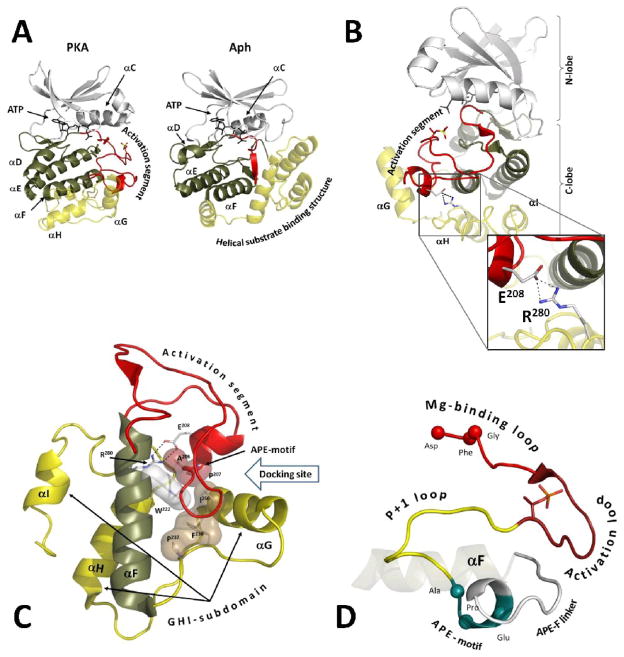
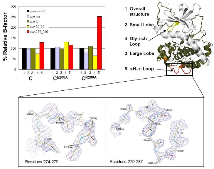
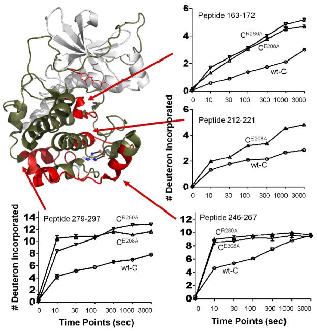
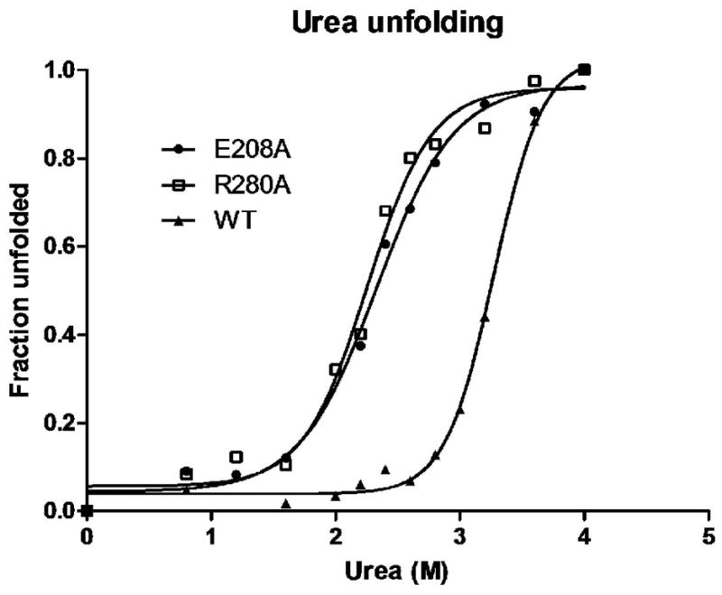
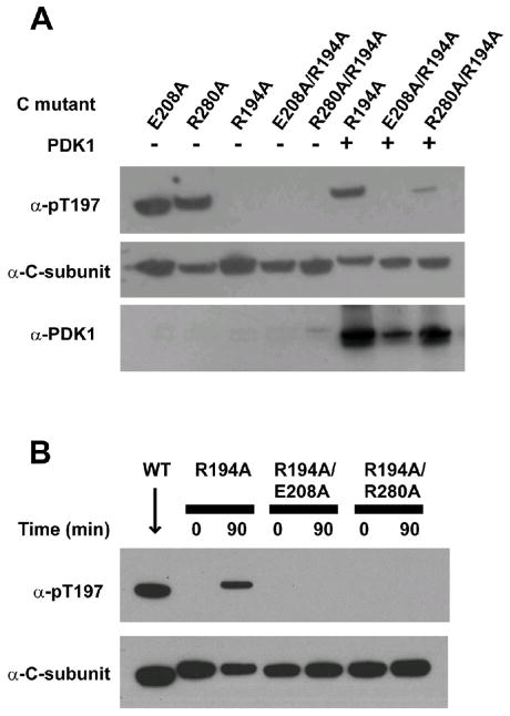
Eukaryotic protein kinases (EPKs) feature two coevolved structural segments, the Activation segment, which starts with the Asp-Phe-Gly (DFG) and ends with the Ala-Pro-Glu (APE) motifs, and the helical GHI subdomain that comprises αG-αH-αI helices. Eukaryotic-like kinases have a much shorter Activation segment and lack the GHI subdomain. They thus lack the conserved salt bridge interaction between the APE Glu and an Arg from the GHI subdomain, a hallmark signature of EPKs. Although the conservation of this salt bridge in EPKs is well known and its implication in diseases has been illustrated by polymorphism analysis, its function has not been carefully studied. In this work, we use murine cAMP-dependent protein kinase (protein kinase A) as the model enzyme (Glu208 and Arg280) to examine the role of these two residues. We showed that Ala replacement of either residue caused a 40- to 120-fold decrease in catalytic efficiency of the enzyme due to an increase in K(m)(ATP) and a decrease in k(cat). Crystal structures, as well as solution studies, also demonstrate that this ion pair contributes to the hydrophobic network and stability of the enzyme. We show that mutation of either Glu or Arg to Ala renders both mutant proteins less effective substrates for upstream kinase phosphoinositide-dependent kinase 1. We propose that the Glu208-Arg280 pair serves as a center hub of connectivity between these two structurally conserved elements in EPKs. Mutations of either residue disrupt communication not only between the two segments but also within the rest of the molecule, leading to altered catalytic activity and enzyme regulation.
Copyright © 2011 Elsevier Ltd. All rights reserved.
Conflict of interest statement
Figures

Similar articles
-
Global consequences of activation loop phosphorylation on protein kinase A.J Biol Chem. 2010 Feb 5;285(6):3825-3832. doi: 10.1074/jbc.M109.061820. Epub 2009 Dec 4. J Biol Chem. 2010. PMID: 19965870 Free PMC article.
-
Evolution of the eukaryotic protein kinases as dynamic molecular switches.Philos Trans R Soc Lond B Biol Sci. 2012 Sep 19;367(1602):2517-28. doi: 10.1098/rstb.2012.0054. Philos Trans R Soc Lond B Biol Sci. 2012. PMID: 22889904 Free PMC article. Review.
-
The "catalytic" triad of isocitrate dehydrogenase kinase/phosphatase from E. coli and its relationship with that found in eukaryotic protein kinases.Biochemistry. 2001 Mar 13;40(10):3047-55. doi: 10.1021/bi001713x. Biochemistry. 2001. PMID: 11258918
-
Catalytic Subunit of PKA as a Prototype of the Eukaryotic Protein Kinase Family.Biochemistry (Mosc). 2020 Apr;85(4):409-424. doi: 10.1134/S0006297920040021. Biochemistry (Mosc). 2020. PMID: 32569549 Review.
-
Functional Role of Histidine in the Conserved His-x-Asp Motif in the Catalytic Core of Protein Kinases.Sci Rep. 2015 May 11;5:10115. doi: 10.1038/srep10115. Sci Rep. 2015. PMID: 25960268 Free PMC article.
Cited by
-
Conformational equilibrium of N-myristoylated cAMP-dependent protein kinase A by molecular dynamics simulations.Biochemistry. 2012 Dec 21;51(51):10186-96. doi: 10.1021/bi301279f. Epub 2012 Dec 12. Biochemistry. 2012. PMID: 23205665 Free PMC article.
-
The dimer-dependent catalytic activity of RAF family kinases is revealed through characterizing their oncogenic mutants.Oncogene. 2018 Oct;37(43):5719-5734. doi: 10.1038/s41388-018-0365-2. Epub 2018 Jun 21. Oncogene. 2018. PMID: 29930381 Free PMC article.
-
Direct phosphorylation of HY5 by SPA kinases to regulate photomorphogenesis in Arabidopsis.New Phytol. 2021 Jun;230(6):2311-2326. doi: 10.1111/nph.17332. Epub 2021 Apr 6. New Phytol. 2021. PMID: 33686674 Free PMC article.
-
Role of key salt bridges in thermostability of G. thermodenitrificans EstGtA2: distinctive patterns within the new bacterial lipolytic enzyme subfamily XIII.2 [corrected].PLoS One. 2013 Oct 8;8(10):e76675. doi: 10.1371/journal.pone.0076675. eCollection 2013. PLoS One. 2013. PMID: 24116134 Free PMC article.
-
Global connectivity of hub residues in Oncoprotein structures encodes genetic factors dictating personalized drug response to targeted Cancer therapy.Sci Rep. 2014 Dec 3;4:7294. doi: 10.1038/srep07294. Sci Rep. 2014. PMID: 25465236 Free PMC article.
References
-
- Kannan N, Neuwald AF. Did protein kinase regulatory mechanisms evolve through elaboration of a simple structural component? J Mol Biol. 2005;351:956–72. - PubMed
-
- Diskar M, Zenn HM, Kaupisch A, Kaufholz M, Brockmeyer S, Sohmen D, Berrera M, Zaccolo M, Boshart M, Herberg FW, Prinz A. Regulation of cAMP-dependent protein kinases: the human protein kinase X (PrKX) reveals the role of the catalytic subunit alphaH-alphaI loop. J Biol Chem. 2010;285:35910–8. - PMC - PubMed
-
- Kim C, Cheng CY, Saldanha SA, Taylor SS. PKA-I holoenzyme structure reveals a mechanism for cAMP-dependent activation. Cell. 2007;130:1032–43. - PubMed
Publication types
MeSH terms
Substances
Associated data
- Actions
- Actions
Grants and funding
LinkOut - more resources
Full Text Sources
Molecular Biology Databases
Research Materials
Miscellaneous
