. 2012 May;9(5):812-20.
doi: 10.1016/j.hrthm.2011.11.055. Epub 2011 Dec 2.

BIN1 is reduced and Cav1.2 trafficking is impaired in human failing cardiomyocytes

Affiliations

BIN1 is reduced and Cav1.2 trafficking is impaired in human failing cardiomyocytes

Ting-Ting Hong et al. Heart Rhythm. 2012 May.

Abstract

Background: Heart failure is a growing epidemic, and a typical aspect of heart failure pathophysiology is altered calcium transients. Normal cardiac calcium transients are initiated by Cav1.2 channels at cardiac T tubules. Bridging integrator 1 (BIN1) is a membrane scaffolding protein that causes Cav1.2 to traffic to T tubules in healthy hearts. The mechanisms of Cav1.2 trafficking in heart failure are not known.

Objective: To study BIN1 expression and its effect on Cav1.2 trafficking in failing hearts.

Methods: Intact myocardium and freshly isolated cardiomyocytes from nonfailing and end-stage failing human hearts were used to study BIN1 expression and Cav1.2 localization. To confirm Cav1.2 surface expression dependence on BIN1, patch-clamp recordings were performed of Cav1.2 current in cell lines with and without trafficking-competent BIN1. Also, in adult mouse cardiomyocytes, surface Cav1.2 and calcium transients were studied after small hairpin RNA-mediated knockdown of BIN1. For a functional readout in intact heart, calcium transients and cardiac contractility were analyzed in a zebrafish model with morpholino-mediated knockdown of BIN1.

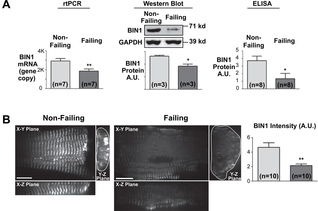
Results: BIN1 expression is significantly decreased in failing cardiomyocytes at both mRNA (30% down) and protein (36% down) levels. Peripheral Cav1.2 is reduced to 42% by imaging, and a biochemical T-tubule fraction of Cav1.2 is reduced to 68%. The total calcium current is reduced to 41% in a cell line expressing a nontrafficking BIN1 mutant. In mouse cardiomyocytes, BIN1 knockdown decreases surface Cav1.2 and impairs calcium transients. In zebrafish hearts, BIN1 knockdown causes a 75% reduction in calcium transients and severe ventricular contractile dysfunction.

Conclusions: The data indicate that BIN1 is significantly reduced in human heart failure, and this reduction impairs Cav1.2 trafficking, calcium transients, and contractility.


Conflict of interest statement

Conflicts of interests: None

Figures

Figure 1
Figure 1. Failing cardiomyocytes express lower BIN1
(A) Quantitative RT-PCR analysis (left), semi-quantitative western blotting and quantitative ELISA indicate significant reduction of BIN1 expression in failing human hearts. (B) Confocal images (100x) of isolated human cardiomyocytes stained with mouse anti-BIN1 antibody indicate decreased expression of BIN1. (*, P<0.05; **, P<0.01)
Figure 2
Figure 2. Intracellular Cav1.2 distribution in failing human cardiomyocytes
(A) Two dimensional frame view of confocal images (100x) of Cav1.2 immunofluorescence in human non-failing (left panel) and failing cardiomyocytes (right panel). (B) Volume view of intracellular Cav1.2 distribution reconstructed from a stack of 100x confocal image frames acquired at a z-step of 0.1 μm. The Z-axis cross section volume views of subsection cardiomyocytes were generated from the original z-stack and shown in the left panel (top, representative non-failing cardiomyocyte; bottom, representative failing cardiomyocyte). The top right cardiomyocyte schematic illustrates the imaging frames (green), the viewing sections (red frames), as well as the analysis of the peripheral Cav1.2 (blue area). Cortical Cav1.2 at the cell periphery within 2 μm of cell edges (blue area indicated in the schematic) were quantified and normalized to total cellular Cav1.2 and are presented in the bottom right panel. Peripheral Cav1.2 fluorescent signal is significantly reduced in failing cardiomyocytes. (***, P<0.001)
Figure 3
Figure 3. Reduced T-tubule abundance of Cav1.2 in failing human myocardium
(A) Western blot of protein lysates prepared from human myocardium indicates no significant change of Cav1.2 protein expression level in failing myocardium. (B) A sucrose gradient was generated for isolation of T-tubule membranes from the microsomes prepared from human myocardium. As indicated, fractions recovered from 32%/35% interface are enriched with T-tubule localized proteins like Cav1.2 and NCX1. In the same T-tubule fraction prepared from failing myocardium, Cav1.2 expression is significantly reduced whereas NCX1 expression is preserved. T-tubule Cav1.2 protein level is normalized to NCX1 and presented in the right panel. (n=3 hearts, P<0.05)
Figure 4
Figure 4. BIN1 level controls surface availability of functional Cav1.2 channels
(A) Surface biotinylation indicate that reduced BIN1 expression following lentiviral mediated shRNA knockdown (left) causes less surface expression of Cav1.2 in adult mouse cardiomyocytes. (B) Exogenous BIN1 rescues Cav1.2 trafficking. In three-day cultured cardiomyocytes (compared to freshly dissociated) BIN1 expression is decreased and Cav1.2 is internalized. Introduction of V5-tagged exogenous BIN1 (indicated as anti-V5 staining) by lentiviral construct normalizes BIN1 level and rescues Cav1.2 surface expression. (C) Non Cav1.2 targeting BIN1 mutant (BIN1-BAR*) decreases full length BIN1 induced surface Cav1.2 current in HEK 293 cells (n=9). The half maximal activation voltage (V1/2) remains the same. (*, P<0.05; **, P<0.01; ***, P<0.001)
Figure 5
Figure 5. BIN1 knockdown delays calcium transient in adult mouse cardiomyocytes
BIN1 knockdown by shRNA (indicated in Figure 4A) delays calcium transient development in adult mouse cardiomyocytes. Average time to half maximal signal (T1/2max) is presented in the bar graph (bottom). (**, P<0.01)
Figure 6
Figure 6. BIN1 knockdown diminishes cardiac calcium transient in zebrafish
Calcium transients are decreased after bin1 morpholino injection in zebrafish. Top panel includes representative cardiac calcium transients in whole zebrafish hearts 70 hpf with control morpholino (top) or bin1 morpholino (bottom). Baseline fluorescent (F0) and maximal fluorescent (Fmax) are indicated. The amplitude of calcium transients (ΔF/F0) are averaged and presented in the bottom panel. (***, P<0.001)
Figure 7
Figure 7. Reduced BIN1 impairs Cav1.2 trafficking and calcium transient regulation
Model of Cav1.2 trafficking and calcium transient regulation in non-failing and failing cardiomyocytes.

Comment in

References

    1. Lloyd-Jones D, Adams RJ, Brown TM, et al. Heart Disease and Stroke Statistics--2010 Update. A Report From the American Heart Association. Circulation. 2010;121:e46–e215. - PubMed
    1. Bauce B, Nava A, Beffagna G, et al. Multiple mutations in desmosomal proteins encoding genes in arrhythmogenic right ventricular cardiomyopathy/dysplasia. Heart Rhythm. 2010;7:22–29. - PubMed
    1. Mohler PJ, Schott JJ, Gramolini AO, et al. Ankyrin-B mutation causes type 4 long-QT cardiac arrhythmia and sudden cardiac death. Nature. 2003;421:634–639. - PubMed
    1. Mohler PJ, Rivolta I, Napolitano C, et al. Nav1.5 E1053K mutation causing Brugada syndrome blocks binding to ankyrin-G and expression of Nav1.5 on the surface of cardiomyocytes. Proc Natl Acad Sci U S A. 2004;101:17533–17538. - PMC - PubMed
    1. Frost A, Unger VM, De Camilli P. The BAR domain superfamily: membrane-molding macromolecules. Cell. 2009;137:191–196. - PMC - PubMed