. 2012 Mar 15;119(11):2533-44.
doi: 10.1182/blood-2011-07-367334. Epub 2011 Dec 2.

Human FcγRIIA induces anaphylactic and allergic reactions

Affiliations

Human FcγRIIA induces anaphylactic and allergic reactions

Friederike Jönsson et al. Blood. .

Abstract

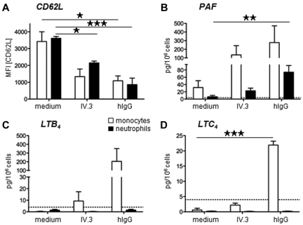
IgE and IgE receptors (FcεRI) are well-known inducers of allergy. We recently found in mice that active systemic anaphylaxis depends on IgG and IgG receptors (FcγRIIIA and FcγRIV) expressed by neutrophils, rather than on IgE and FcεRI expressed by mast cells and basophils. In humans, neutrophils, mast cells, basophils, and eosinophils do not express FcγRIIIA or FcγRIV, but FcγRIIA. We therefore investigated the possible role of FcγRIIA in allergy by generating novel FcγRIIA-transgenic mice, in which various models of allergic reactions induced by IgG could be studied. In mice, FcγRIIA was sufficient to trigger active and passive anaphylaxis, and airway inflammation in vivo. Blocking FcγRIIA in vivo abolished these reactions. We identified mast cells to be responsible for FcγRIIA-dependent passive cutaneous anaphylaxis, and monocytes/macrophages and neutrophils to be responsible for FcγRIIA-dependent passive systemic anaphylaxis. Supporting these findings, human mast cells, monocytes and neutrophils produced anaphylactogenic mediators after FcγRIIA engagement. IgG and FcγRIIA may therefore contribute to allergic and anaphylactic reactions in humans.


Figures

Figure 1
Figure 1
FcγRIIA can induce active systemic anaphylaxis. (A) Representative histogram plots of human FcR expression on human blood neutrophils. (B) Representative histogram plots of anti-FLAG mAb (top panel) or preformed mouse polyclonal IgG-immune complexes (bottom panel) binding to CHO transfectants expressing the indicated FLAG-tagged human polymorphic variants of FcγRIIA (H131 or R131) and FcγRIIIB (NA1, NA2, or SH). (C) Representative expression of FcγRIIA on blood and peritoneal cells from 3KOIIA mice (open histograms) or nontransgenic 3KO littermate controls (filled histograms): T cells (CD3+), B cells (CD19+), NK cells (DX5+/NK1.1+), neutrophils (Gr1hi/CD11b+), eosinophils (Gr1int/SiglecF+), basophils (IgE+/DX5+), and monocytes/macrophages (CD11b+/Gr1). (D) Representative expression of FcγRIIA (open histograms) on human blood cells: T cells (CD3+), B cells (CD19+), NK cells (CD56+), neutrophils (CD24+), eosinophils (CCR3+/CDw125+), basophils (FcϵRI+/CD203c+), and monocytes (CD14+); or isotype control (closed histograms). (E-G) Indicated mice were immunized with BSA, (E) in Freunds adjuvant, or (F-G) in alum, challenged with BSA and central temperatures and survival rates were monitored. (E) ASA in FcRγ−/− (n = 7) and FcRγ−/−IIA mice (n = 5). (F) ASA in FcRγ−/− (n = 4) and FcRγ−/−IIA (n = 5) mice. (G) ASA in FcRγ−/−IIA mice injected twice with anti-FcγRIIA mAb IV.3 (n = 5) or not (n = 6), before BSA-challenge. FcRγ−/− mice were used as controls (n = 7). (E-G) Data are represented as mean ± SEM. (A-G) Data are representative of at least 2 independent experiments (*P < .05; **P < .01; ***P < .001).
Figure 2
Figure 2
In vivo aggregation of FcγRIIA induces passive systemic anaphylaxis. (A,C-D) Indicated FcγRIIA-transgenic mice were injected with 50 μg (gray symbols) or 150 μg (black symbols) of mAb IV.3, and central temperatures were monitored (n = 3). Nontransgenic littermates injected with 150 μg mAb IV.3 were used as controls (open symbols, n = 3). (A) FcRγ−/−, (C) 3KO, (D) 5KO backgrounds. (B) Schematic representation of Fc receptors expressed in the different mouse models used in this study. (E-F) Mice were injected with indicated preformed mouse IC and central temperatures were monitored. Gray symbols indicate mice injected with 50 μg of mAb IV.3 24 hours before challenge (n = 4). Top panel (E) n = 5, (F) n = 4. Bottom panel (E) 3KO or 3KOIIA n = 3, (F) 3KO n = 3, 3KOIIA+iso n = 4. (A,C-F) Data are represented as mean ± SEM and are representative of at least 2 independent experiments (**P < .01; ***P < .001).
Figure 3
Figure 3
Neutrophils and monocytes/macrophages are necessary for FcγRIIA-dependent PSA. (A-G) Indicated mice were injected with preformed polyclonal IgG-IC (mouse anti-GPI antiserum plus GPI), and central temperatures were monitored. PSA in FcγRIIA transgenic mice injected with (A) anti-Gr1 mAbs (n = 8) or isotype (ISO) control (n = 3), (B) anti–Ly-6G mAbs (n = 4) or untreated control (n = 4), (C) anti-CD200R3 mAbs (n = 3) or untreated control (n = 3). Nontransgenic littermates were used as controls (A, n = 2; B, n = 4; C, n = 3). (D) PSA in Wsh3KOIIA mice and 3KOIIA mice (n = 3). Nontransgenic littermate controls 3KO (n = 3) and Wsh3KO mice (n = 4) were used as controls. (E-F) PSA in 3KOIIA mice injected with (E) PBS liposomes (PBS lipo) or clodronate liposomes (toxic lipo; n = 3), (F) Gadolinium chloride (GdCl3) or not (n = 4). Nontransgenic littermates were used as controls (E, n = 2; F: n = 4). (G) PSA in 3KOIIA mice left untreated (n = 7), or injected with anti-Gr1 mAbs (n = 5), toxic liposomes (n = 6) or anti-Gr1 mAbs plus toxic liposomes (n = 6). Data are a compilation of 2 experiments. 3KO served as negative control (n = 2). Statistical significances are indicated among 3KOIIA groups. (H) mAb IV.3-induced PSA in indicated mice injected with anti-Gr1 mAbs, toxic liposomes, or left untreated (n = 3). (A-H) Data are represented as mean ± SEM, and (A-F,H) are representative of at least 2 independent experiments (*P < .05; **P < .01; ***P < .001).
Figure 4
Figure 4
Human neutrophils and monocytes produce anaphylactogenic mediators. (A-D) Purified human monocytes or neutrophils were incubated in vitro with heat-aggregated human IgG or anti-FcγRIIA mAb IV.3, and (A) CD62L expression, (B) PAF, (C) LTB4, and (D) LTC4 production are represented. Mean results from the analysis of 3 normal donors are represented (*P < .05; **P < .01; ***P < .001).
Figure 5
Figure 5
Mast cells are mandatory for FcγRIIA-dependent passive cutaneous anaphylaxis. (A-C) Mice were injected intradermally with indicated reagents and intravenously with Evans blue. Quantification of Evans blue extracted from skin tissue is represented. (A-B) PCA in 5KO (open symbols: A, n = 4; B, n = 3), 5KOIIA (black symbols: A, n = 3, B, n = 3), or 5KOIIA mice preinjected (A) once or (B) twice with 50 μg mAb IV.3 (gray symbols, n = 4). (C) PCA in 3KOIIA mice (black symbols, n = 4), in Wsh3KOIIA (gray symbols, n = 3), and as controls in Wsh3KO (open symbols, n = 2). NB: 2 points are represented per mouse, as each mouse was injected on 2 different sites with Ag and with IC. (D) Representative expression of FcγRIIA on peritoneal mast cells (c-kit+/IgE+) from 3KOIIA mice (open histograms) or 3KO littermate controls (filled histograms). (E-F) Peritoneal cells from 3KO (white bars) and 3KOIIA (black bars) mice were stimulated with indicated reagents. (E) The percentage of degranulated mast cells (a minimum of 200 cells were counted per experimental point), and (F) histamine content in the supernatant of each experimental point are represented. (A-C) Data are represented as single measured points, and mean ± SEM. (A-F) Data are representative of at least 2 independent experiments (*P < .05; ***P < .001).
Figure 6
Figure 6
FcγRIIA activates mouse and human mast cells in vitro. (A) Representative expression of FcγRIIA on PCMCs, FSMCs, and BMMCs from 3KOIIA (open histogram) and 3KO mice (filled histogram). (B) PCMCs from indicated mice were incubated with indicated preformed mouse IgG-IC (αOVA: anti-OVA mAb; αGPI: polyclonal anti-GPI antiserum; open histograms) or not (filled histograms). Binding of ICs was detected by staining with F(ab′)2 GAM-PE. (C) Calcium fluxes in PCMCs from 3KO or 3KOIIA mice incubated with indicated IC (black curves) or Ag alone (gray curves). Ionomycin was used as control. (D) Western blot analysis of PCMC lysates after stimulation with indicated reagents for different periods of time. PCMCs sensitized overnight with IgE anti-DNP and challenged with DNP-HSA for 3 minutes served as positive controls. Actin was used as a loading control. FcγRIIA was used as a genotype control (reprobe after pERK1/2 staining). (E-F) Mediator release by PCMCs from 3KO (open bars) and 3KOIIA (black bars) mice challenged with indicated reagents. PCMCs sensitized overnight with IgE anti-DNP and challenged with DNP-HSA served as positive controls. NB: GPI+αGPI correspond to ICs made of GPI and polyclonal anti-GPI antiserum in panel E, and to ICs made of GPI and IgG purified from anti-GPI antiserum in panel F. (G) Representative histogram plots of human FcR expression on human SMCs. (H) Percentage of β-hexosaminidase release and quantification of histamine release by human SMCs incubated with anti-FcϵRI mAb or with preformed complexes of mAb IV.3 and GAM. (E-F,H) Data are represented as mean ± SEM. (A-H) Data are representative from at least 2 independent experiments (*P < .05; **P < .01; ***P < .001).
Figure 7
Figure 7
FcγRIIA can induce acute airway inflammation. (A-B) Sections of (A) human lung or (B) lung from 3KO or 3KOIIA mice, stained with Hematoxilin (blue) and anti-FcγRIIA rabbit antiserum (red). (C) Representative expression of FcγRIIA on mouse alveolar macrophages (CD11c+/Gr1) from 3KOIIA (open histogram) and 3KO (filled histogram) mice. (D) F(ab′)2 DAR-FITC staining of WT or FcγRIIA-expressing CHO transfectants incubated with preformed ICs made of OVA and rabbit anti-OVA antiserum (open histograms) or not (filled histograms). (E-F) Representative density plots of CD45+ BAL cells from indicated mice (E) left untreated or (F) injected with antigen intravenously and antiserum intranasally. Cell types were discriminated as alveolar macrophages (CD11c+/Gr1int, oval gate) and neutrophils (CD11c/Gr1hi, rectangular gate). (G-J) Time course of (G) cell counts, (H) MPO level, (I) hemorrhage score, and (J) KC levels in BAL from indicated mice after injection with antigen intravenously and antiserum intranasally (n ≥ 3). (K) KC secretion or (L) MIP-1α secretion by purified alveolar macrophages from indicated mice incubated ex vivo on plate-bound rabbit IgG-ICs (OVA–anti-OVA) or IV.3 mAb. (G-L) Data are represented as mean ± SEM. (A-L) Data are representative from at least 2 independent experiments, and (J) data are a compilation of 2 experiments (*P < .05; **P < .01; ***P < .001).

Comment in

  • Human FcγRIIA at center stage.
    Kawakami T. Kawakami T. Blood. 2012 Mar 15;119(11):2432-3. doi: 10.1182/blood-2012-01-397786. Blood. 2012. PMID: 22422810 No abstract available.

Similar articles

Cited by

References

    1. Jönsson F, Mancardi DA, Kita Y, et al. Mouse and human neutrophils induce anaphylaxis. J Clin Invest. 2011;121(4):1484–1496. - PMC - PubMed
    1. Daëron M. Fc receptor biology. Annu Rev Immunol. 1997;15:203–234. - PubMed
    1. Wershil BK, Mekori YA, Murakami T, Galli SJ. 125I-fibrin deposition in IgE-dependent immediate hypersensitivity reactions in mouse skin. Demonstration of the role of mast cells using genetically mast cell-deficient mice locally reconstituted with cultured mast cells. J Immunol. 1987;139(8):2605–2614. - PubMed
    1. Arimura A, Nagata M, Takeuchi M, Watanabe A, Nakamura K, Harada M. Active and passive cutaneous anaphylaxis in WBB6F1 mouse, a mast cell-deficient strain. Immunol Invest. 1990;19(3):227–233. - PubMed
    1. Arimura A, Nagata M, Watanabe A, Nakamura K, Takeuchi M, Harada M. Production of active and passive anaphylactic shock in the WBB6F1 mouse, a mast cell-deficient strain. Experientia. 1990;46(7):739–742. - PubMed

Publication types

MeSH terms