. 2012 Mar 15;119(11):2644-56.
doi: 10.1182/blood-2011-08-371971. Epub 2011 Dec 2.

Adoptive immunotherapy with unselected or EBV-specific T cells for biopsy-proven EBV+ lymphomas after allogeneic hematopoietic cell transplantation

Affiliations

Adoptive immunotherapy with unselected or EBV-specific T cells for biopsy-proven EBV+ lymphomas after allogeneic hematopoietic cell transplantation

Ekaterina Doubrovina et al. Blood. .

Abstract

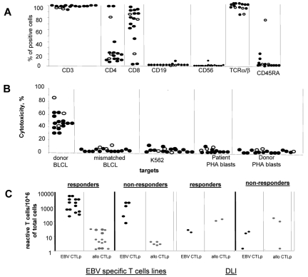
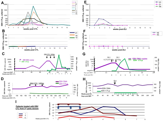
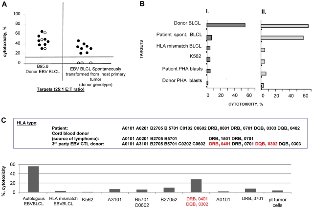
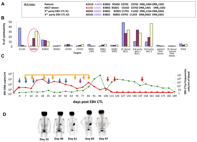
We evaluated HLA-compatible donor leukocyte infusions (DLIs) and HLA-compatible or HLA-disparate EBV-specific T cells (EBV-CTLs) in 49 hematopoietic cell transplantation recipients with biopsy-proven EBV-lymphoproliferative disease (EBV-LPD). DLIs and EBV-CTLs each induced durable complete or partial remissions in 73% and 68% of treated patients including 74% and 72% of patients surviving ≥ 8 days after infusion, respectively. Reversible acute GVHD occurred in recipients of DLIs (17%) but not EBV-CTLs. The probability of complete response was significantly lower among patients with multiorgan involvement. In responders, DLIs and EBV-CTLs regularly induced exponential increases in EBV-specific CTL precursor (EBV-CTLp) frequencies within 7-14 days, with subsequent clearance of EBV viremia and resolution of disease. In nonresponders, EBV-CTLps did not increase and EBV viremia persisted. Treatment failures were correlated with impaired T-cell recognition of tumor targets. Either donor-derived EBV-CTLs that had been sensitized with autologous BLCLs transformed by EBV strain B95.8 could not lyse spontaneous donor-derived EBV-transformed BLCLs expanded from the patient's blood or biopsied tumor or they failed to lyse their targets because they were selectively restricted by HLA alleles not shared by the EBV-LPD. Therefore, either unselected DLIs or EBV-specific CTLs can eradicate both untreated and Rituxan-resistant lymphomatous EBV-LPD, with failures ascribable to impaired T-cell recognition of tumor-associated viral antigens or their presenting HLA alleles.


Figures

Figure 1
Figure 1
Characterization of the EBV specific T-cell lines. (A) Phenotype of the EBV specific T-cell lines used for the treatment of EBV-LPD in patients who responded (closed symbols) and did not respond (open symbols) to the T-cell therapy. All EBV-CTL lines infused contained more than 90% of CD3+ cells with predominance of CD8+ T cells. However, 3 CTL lines contained predominantly CD4+ T cells. Infusion of the CD4+ T cell–predominant lines achieved CR in 2 of the 3 patients infused. All T-cell lines were equally depleted of NK and B cells. (B) Cytotoxic activity of the EBV-CTL lines used for the treatment of EBV-LPD of those patients who responded (closed symbols) and those who did not respond (open symbols) to the T-cell therapy. (C) Frequencies of EBV-specific T cells (black symbols) and alloreactive (gray symbols) detected by limiting dilution analysis in EBV-specific T-cell lines and DLI products before their use for the treatment of EBV-LPD demonstrate higher frequencies of EBV-specific T cells and lower frequencies of alloreactive T cells in the EBV-specific T-cell lines than in the unstimulated donor leukocytes. There were no differences in the frequencies of EBV-specific and alloreactive cells between the cell products used in responders and nonresponders. (D) All EBV-CTL lines infused exhibited exclusively EBV-specific cytotoxicity without any activity against recipient PHA-activated blasts, mismatched EBV-BLCLs, and K562 (a target for NK cells).
Figure 2
Figure 2
Monitoring of EBV-specific T cells in the circulation of patients with EBV-LPD treated with EBV-specific T cells or unstimulated DLIs. (A) Frequencies of EBV-specific T cells observed over a period of 12 weeks after initiation of T-cell therapy in the peripheral blood of patients who responded to treatment with EBV-specific T cells obtained from their stem cell donors (n = 8 patients evaluated). (B) Frequencies of EBV-specific T cells observed in peripheral blood of the patients with EBV-LPD who did not respond to treatment with EBV-specific T cells (n = 3) generated from PBMCs of their stem cell donors. (C) Monitoring of the EBV DNA levels (purple line) and EBV-CTLps (green line) of patient 15, who responded to treatment with EBV-specific T cells derived from the stem cell donor. EBV-CTLs were infused weekly for 3 consecutive weeks at doses of 1 × 10⁶ cells/kg/infusion. After infusions of T cells, EBV-CTLps increased in frequency. Concurrently, EBV DNA levels in the peripheral blood of the patient decreased. (D) Monitoring of the EBV DNA levels (purple line) and EBV-CTLps (green line) of patient 41 who did not respond to treatment with EBV-specific T cells derived from the stem cell donor. EBV-CTLs were infused weekly for 3 consecutive weeks at doses of 1 × 10⁶ cells/kg/infusion. No increase in EBV-CTLp frequency was observed over the period of 6 weeks of observation and the levels of EBV DNA remained high. (E) EBV-CTLps in patients who responded to therapy with DLIs from their HLA-matched stem cell donors (n = 3 patients evaluated). (F) EBV-CTLps in patients who did not respond to the therapy with DLIs (patients 43 and 45). (G-H) EBV DNA levels (purple line) and EBV-specific CTLps (green line) in the peripheral blood of patient 21, who responded to DLI treatment (G) and patient 45, who did not respond to the infusion of DLIs (H). (J) Monitoring of T-cell responses in the peripheral blood of patients with EBV-LPD who received EBV-specific T cells generated from third-party healthy donors (n = 5). The arrows indicate the times of the T-cell infusions for each of 3 patients treated and are presented in the same color as the line demonstrating the CTLps frequencies. Patients 1 and 6 achieved CR; patient 13 did not respond and died of PD.
Figure 3
Figure 3
Basis for nonresponse: donor-derived EBV-specific T cells that failed to induce a clinical response did not exhibit cytotoxic activity against the spontaneously transformed EBV-BLCLs generated from the patient's tissues. (A) Cytotoxicity of donor EBV–specific CTLs. There were 8 responders (closed symbols) and 3 nonresponders (open symbols; n = 11) sensitized with autologous B95.8-transformed B cells against EBV B95.8–transformed donor–derived autologous BLCL and against donor-type spontaneous EBV-transformed BLCLs cultured from the patient blood or EBV-LPD. The B95.8-sensitized EBV-specific CTLs used in nonresponding patients lysed EBV B95.8 transformed BLCL but did not lyse the spontaneous EBV transformants of donor origin cultured from the patient. (B) Cytotoxicity of donor-derived EBV-CTLs stimulated with spontaneously transformed EBV-BLCLs of donor origin cultured from the patient. The same T cells as in panel A were stimulated in vitro with the spontaneously transformed EBV-BLCLs and were able to kill both the stimulating B-cell line and the donor-derived B95.8 transformant. These data confirm that the EBV-LPD is sensitive to lysis by donor-derived EBV-CTLs if the T cells are sensitized with the endogenous strain of EBV. (C) HLA restriction of EBV-CTLs generated from an HLA haplotype–matched donor for patient 13, who developed an EBV lymphoma in cord blood–derived B cells; these EBV-CTLs were selectively restricted by an HLA DRB, 0401, not shared by the cord blood cells and did not lyse cord blood donor-derived spontaneous EBV-BLCL generated from the tumor.
Figure 4
Figure 4
Basis for nonresponse: donor-derived EBV-specific T cells did not recognize host origin EBV-LPD (patient 8). (A) HLA type of the stem cell donor (mother), third-party EBV-CTLs donor 1, and third-party EBV-CTL donor 2. (B) HLA-restriction analysis of EBV-specific T cells generated from the HSCT donor (mother; blue bars) or from the third-party donor 1 (brown bars) or third-party donor 2 (yellow bars) tested in a Cr51-release assay against a panel of allogeneic EBV-BLCLs, each matching 1 HLA allele of each of the T-cell donors' HLA types. (C) Monitoring of the EBV DNA levels (red line) and EBV-specific T cells (green line) after infusions of EBV-CTLs derived from the stem cell donor (blue arrows), third-party donor 1 (purple arrow), and third-party donor 2 (red arrows) at the doses of 1 × 10⁶ cells/kg/infusion. Treatment with Rituxan (375mg/m2; orange arrows), as well as injections of EBV-CTLs derived from the HSCT donor that were restricted by the donor-unique HLA A1101 allele, did not affect high levels EBV DNA, whereas the administration of the EBV-specific T cells from the third-party donors 1 and 2, both restricted by the A2601 HLA allele presented on the EBV+ tumor cells of patient origin, resulted in a rapid decrease of the EBV DNA in the circulation. (D) Sequential PET scans demonstrating no response to the transplant donor's T cells with rapid development of an EBV lymphoma of the gastric wall and adjacent lymph nodes. By 3 weeks after the first infusion of third-party T cells (d97), gastric lymphoma was no longer detected.

Comment in

Similar articles

Cited by

References

    1. Martin PJ, Shulman HM, Schubach WH, et al. Fatal Epstein-Barr-virus-associated proliferation of donor B cells after treatment of acute graft-versus-host disease with a murine anti-T-cell antibody. Ann Intern Med. 1984;101(3):310–315. - PubMed
    1. Peres E, Savasan S, Klein J, Abidi M, Dansey R, Abella E. High fatality rate of Epstein-Barr virus-associated lymphoproliferative disorder occurring after bone marrow transplantation with rabbit antithymocyte globulin conditioning regimens. J Clin Microbiol. 2005;43(7):3540–3543. - PMC - PubMed
    1. Lucas KG, Small TN, Heller G, Dupont B, O'Reilly RJ. The development of cellular immunity to Epstein-Barr virus after allogeneic bone marrow transplantation. Blood. 1996;87(6):2594–2603. - PubMed
    1. Meijer E, Cornelissen JJ. Epstein-Barr virus-associated lymphoproliferative disease after allogeneic haematopoietic stem cell transplantation: molecular monitoring and early treatment of high-risk patients. Curr Opin Hematol. 2008;15(6):576–585. - PubMed
    1. Brunstein CG, Weisdorf DJ, DeFor T, et al. Marked increased risk of Epstein-Barr virus-related complications with the addition of antithymocyte globulin to a nonmyeloablative conditioning prior to unrelated umbilical cord blood transplantation. Blood. 2006;108(8):2874–2880. - PMC - PubMed

Publication types

MeSH terms