

Revisiting the role of hCG: new regulation of the angiogenic factor EG-VEGF and its receptors
- PMID: 22138749
- PMCID: PMC11115148
- DOI: 10.1007/s00018-011-0889-x
Revisiting the role of hCG: new regulation of the angiogenic factor EG-VEGF and its receptors
Abstract
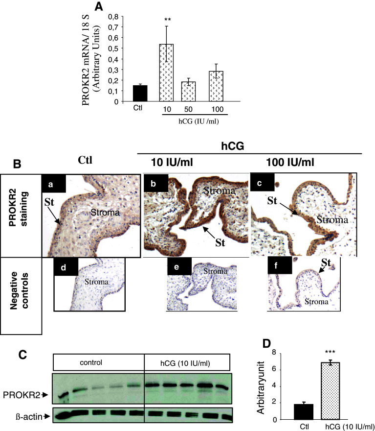
Endocrine gland-derived vascular endothelial growth factor (EG-VEGF) is an angiogenic factor reported to be specific for endocrine tissues, including the placenta. Its biological activity is mediated via two G protein-coupled receptors, prokineticin receptor 1 (PROKR1) and prokineticin receptor 2 (PROKR2). We have recently shown that (i) EG-VEGF expression peaks between the 8th and 11th weeks of gestation, (ii) its mRNA and protein levels are up-regulated by hypoxia, (iii) EG-VEGF is a negative regulator of trophoblast invasion and (iv) its circulating levels are increased in preeclampsia (PE), the most threatening pathology of pregnancy. Here, we investigated the regulation of the expression of EG-VEGF and its receptors by hCG, a key pregnancy hormone that is also deregulated in PE. During the first trimester of pregnancy, hCG and EG-VEGF exhibit the same pattern of expression, suggesting that EG-VEGF is potentially regulated by hCG. Both placental explants (PEX) and primary cultures of trophoblasts from the first trimester of pregnancy were used to investigate this hypothesis. Our results show that (i) LHCGR, the hCG receptor, is expressed both in cyto- and syncytiotrophoblasts, (ii) hCG increases EG-VEGF, PROKR1 and PROKR2 mRNA and protein expression in a dose- and time-dependent manner, (iii) hCG increases the release of EG-VEGF from PEX conditioned media, (iv) hCG effects are transcriptional and post-transcriptional and (v) the hCG effects are mediated by cAMP via cAMP response elements present in the EG-VEGF promoter region. Altogether, these results demonstrate a new role for hCG in the regulation of EG-VEGF and its receptors, an emerging regulatory system in placental development.
Figures


Similar articles
-
EG-VEGF controls placental growth and survival in normal and pathological pregnancies: case of fetal growth restriction (FGR).Cell Mol Life Sci. 2013 Feb;70(3):511-25. doi: 10.1007/s00018-012-1141-z. Epub 2012 Sep 2. Cell Mol Life Sci. 2013. PMID: 22941044 Free PMC article.
-
Expression and oxygen regulation of endocrine gland-derived vascular endothelial growth factor/prokineticin-1 and its receptors in human placenta during early pregnancy.Endocrinology. 2006 Apr;147(4):1675-84. doi: 10.1210/en.2005-0912. Epub 2005 Dec 29. Endocrinology. 2006. PMID: 16384869
-
Molecular characterization of EG-VEGF-mediated angiogenesis: differential effects on microvascular and macrovascular endothelial cells.Mol Biol Cell. 2010 Aug 15;21(16):2832-43. doi: 10.1091/mbc.E10-01-0059. Epub 2010 Jun 29. Mol Biol Cell. 2010. PMID: 20587779 Free PMC article.
-
Prokineticin1 and pregnancy.Ann Endocrinol (Paris). 2016 Jun;77(2):101-4. doi: 10.1016/j.ando.2016.04.014. Epub 2016 May 9. Ann Endocrinol (Paris). 2016. PMID: 27172869 Review.
-
EG-VEGF: a key endocrine factor in placental development.Trends Endocrinol Metab. 2012 Oct;23(10):501-8. doi: 10.1016/j.tem.2012.05.006. Epub 2012 Jun 16. Trends Endocrinol Metab. 2012. PMID: 22709436 Review.
Cited by
-
Placental Development and Pregnancy-Associated Diseases.Matern Fetal Med. 2021 Dec 14;4(1):36-51. doi: 10.1097/FM9.0000000000000134. eCollection 2022 Jan. Matern Fetal Med. 2021. PMID: 40406576 Free PMC article. Review.
-
Genetic resilience to amyloid related cognitive decline.Brain Imaging Behav. 2017 Apr;11(2):401-409. doi: 10.1007/s11682-016-9615-5. Brain Imaging Behav. 2017. PMID: 27743375 Free PMC article.
-
A case study on the potential angiogenic effect of human chorionic gonadotropin hormone in rapid progression and spontaneous regression of metastatic renal cell carcinoma during pregnancy and after surgical abortion.BMC Cancer. 2015 Dec 24;15:1013. doi: 10.1186/s12885-015-2031-1. BMC Cancer. 2015. PMID: 26704433 Free PMC article.
-
Integrative transcriptome meta-analysis reveals widespread sex-biased gene expression at the human fetal-maternal interface.Mol Hum Reprod. 2014 Aug;20(8):810-9. doi: 10.1093/molehr/gau035. Epub 2014 May 27. Mol Hum Reprod. 2014. PMID: 24867328 Free PMC article.
-
Molecular mechanisms of syncytin-1 in tumors and placental development related diseases.Discov Oncol. 2023 Jun 16;14(1):104. doi: 10.1007/s12672-023-00702-6. Discov Oncol. 2023. PMID: 37326913 Free PMC article. Review.
References
-
- Brouillet S, Hoffmann P, Benharouga M, Salomon A, Schaal JP, Feige JJ, Alfaidy N. Molecular characterization of EG-VEGF-mediated angiogenesis: differential effects on microvascular and macrovascular endothelial cells. Mol Biol Cell. 2010;21:2832–2843. doi: 10.1091/mbc.E10-01-0059. - DOI - PMC - PubMed
-
- Battersby S, Critchley HO, Morgan K, Millar RP, Jabbour HN. Expression and regulation of the prokineticins (endocrine gland-derived vascular endothelial growth factor and Bv8) and their receptors in the human endometrium across the menstrual cycle. J Clinical Endocrinol Metabolism. 2004;89:2463–2469. doi: 10.1210/jc.2003-032012. - DOI - PubMed
Publication types
MeSH terms
Substances
LinkOut - more resources
Full Text Sources
Other Literature Sources
