Review
. 2011 Dec 5;10(1):51-65.
doi: 10.1038/nrmicro2675.

Conventional and unconventional mechanisms for capping viral mRNA

Affiliations
Review

Conventional and unconventional mechanisms for capping viral mRNA

Etienne Decroly et al. Nat Rev Microbiol. .

Abstract

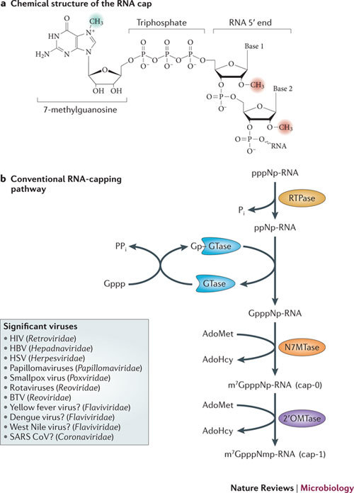
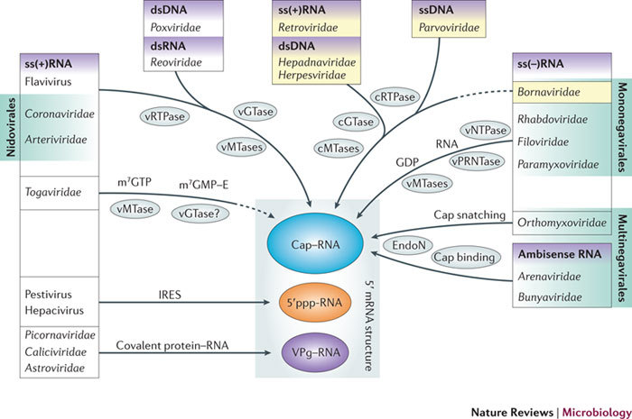
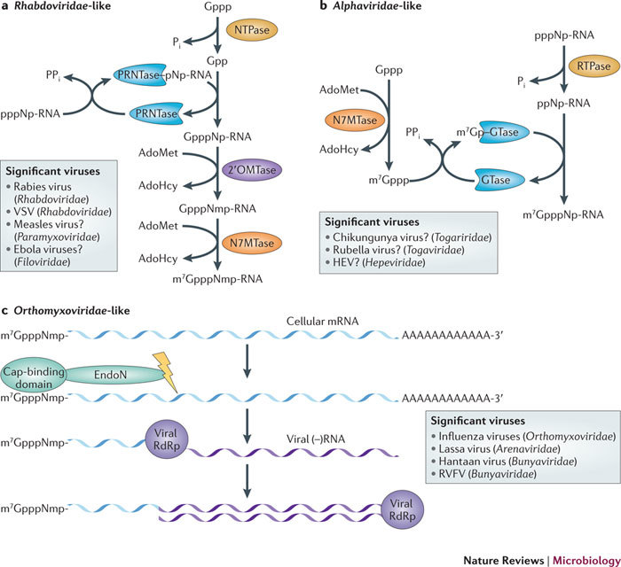
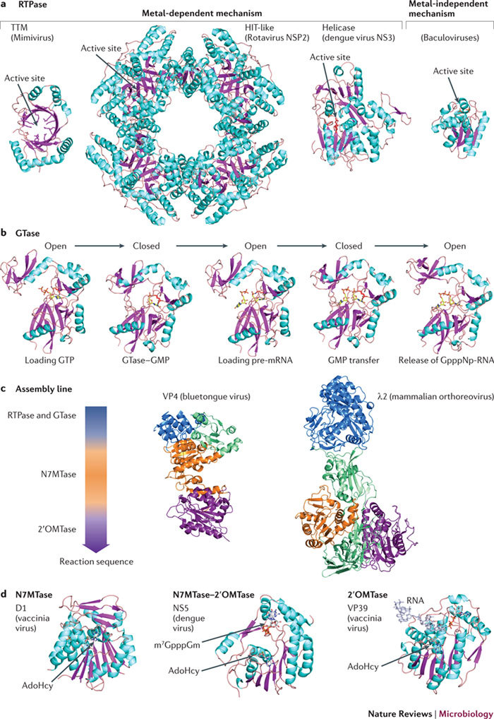
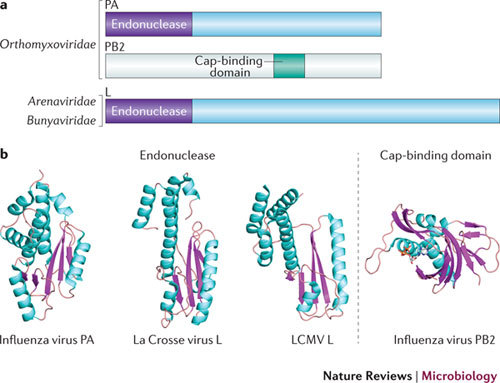
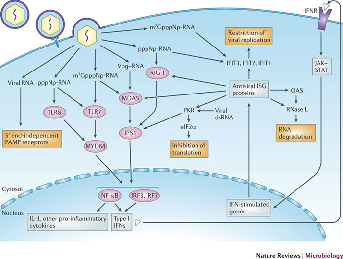
In the eukaryotic cell, capping of mRNA 5' ends is an essential structural modification that allows efficient mRNA translation, directs pre-mRNA splicing and mRNA export from the nucleus, limits mRNA degradation by cellular 5'-3' exonucleases and allows recognition of foreign RNAs (including viral transcripts) as 'non-self'. However, viruses have evolved mechanisms to protect their RNA 5' ends with either a covalently attached peptide or a cap moiety (7-methyl-Gppp, in which p is a phosphate group) that is indistinguishable from cellular mRNA cap structures. Viral RNA caps can be stolen from cellular mRNAs or synthesized using either a host- or virus-encoded capping apparatus, and these capping assemblies exhibit a wide diversity in organization, structure and mechanism. Here, we review the strategies used by viruses of eukaryotic cells to produce functional mRNA 5'-caps and escape innate immunity.


Conflict of interest statement

The authors declare no competing financial interests.

Figures

Figure 1
Figure 1. RNA cap structure and canonical capping mechanisms.
a | The mRNA cap consists of a 7-methylguanosine linked to the 5′ nucleoside of the mRNA chain through a 5′–5′ triphosphate bridge. The methyl group at the N7 position of the guanosine is shaded green, and the 2′-O-methyl groups of the first and second nucleotide residues, forming the cap-1 and the cap-2 structures, respectively, are shaded red. b | The cap-0 structure is formed on nascent RNA chains by the sequential action of three enzymes. First, the RNA triphosphatase (RTPase) hydrolyses the γ-phosphate of the nascent RNA (pppNp-RNA, in which N denotes the first transcribed nucleotide and p denotes a phosphate group) to yield a diphosphate RNA (ppNp-RNA) and inorganic phosphate (Pi). Then, guanylyltransferase (GTase) reacts with the α-phosphate of GTP (Gppp), releasing pyrophosphate (PPi) and forming a covalent enzyme–guanylate intermediate (Gp–GTase). The GTase then transfers the GMP molecule (Gp) to the 5′-diphosphate RNA to create GpppNp-RNA. In the final step, (guanine-N7)-methyltransferase (N7MTase) transfers the methyl group from S-adenosyl-L-methionine (AdoMet) to the cap guanine to form the cap-0 structure, 7-methyl-GpppNp (m7GpppNp), and releases S-adenosyl-L-homocysteine (AdoHcy) as a by-product. The capping reaction is completed by methylation of the ribose-2′-O position of the first nucleotide by the AdoMet-dependent (nucleoside-2′-O)-methyltransferase (2′OMTase), generating the cap-1 structure (m7GpppNm2′-Op). The box contains examples of viruses that acquire their cap structures using the cellular capping machinery or encode their own viral capping machineries that adopt the canonical pathway. Question marks indicate viruses that are likely to follow this conventional pathway. The RNAs capped by viral enzymes are indistinguishable from cellular mRNA and can thus be translated into proteins by the cellular ribosomal machinery. BTV, bluetongue virus; HBV, hepatitis B virus; HSV, herpes simplex viruses; SARS CoV, severe acute respiratory syndrome coronavirus.
Figure 2
Figure 2. RNA 5′ ends in the mammalian-virus world.
Mammalian viruses, with the exception of those from the single-stranded positive-sense RNA (ss(+)RNA) virus genera Pestivirus and Hepacivirus, use strategies to chemically modify their mRNA 5′ ends through either covalent attachment of a protein (VPg for ss(+)RNA viruses from the families Picornaviridae, Caliciviridae and Astroviridae) or covalent attachment of an RNA cap structure (all other viruses). Arrows indicate the type of RNA 5′ end protection that is used by these viral groups, and the enzymes and mechanisms involved are indicated. Dashed arrows indicate a likely but incompletely demonstrated pathway. Viral and cellular proteins are distinguished with the prefixes v and c, respectively. Yellow shading highlights viral groups for which the life cycle includes a nuclear phase in the host cell. This list of viral taxa is non exhaustive and used as an example only. ds, double-stranded; E, enzyme; EndoN, endonuclease; GTase, guanylyltransferase; IRES, internal ribosome entry site; m7, 7-methyl; MTase, methyltransferase; NTPase, nucleotide 5′-triphosphatase; p, phosphate group; PRNTase, polyribonucleotidyl transferase; RTPase, RNA triphosphatase; ss(−)RNA, single-stranded negative-sense RNA.
Figure 3
Figure 3. Unconventional capping pathways.
a | The RNA-capping mechanism of negative-sense RNA ((−)RNA) viruses such as those of the family Rhabdoviridae. The NTPase hydrolyses the γ-phosphate of GTP (Gppp) to yield a GDP (Gpp) and inorganic phosphate (Pi). Polyribonucleotidyl transferase (PRNTase) reacts with the nascent viral RNA (pppNp-RNA; in which N denotes the first transcribed nucleotide), releasing pyrophosphate (PPi) and forming a covalent PRNTase–pNp-RNA intermediate. The PRNTase then transfers the RNA molecule to the GDP to create GpppNp-RNA. (Nucleoside-2′-O)-methyltransferase (2′OMTase) transfers the methyl group from S-adenosyl-L-methionine (AdoMet) to the first nucleotide of the RNA. The capping reaction is then completed by methylation of the cap by the AdoMet-dependent (guanine-N7)-methyltransferase (N7MTase). The box lists examples of viruses that acquire their cap structures using such a capping pathway. Question marks indicate viruses that are likely to follow this conventional pathway. b | The RNA-capping mechanism of positive-sense RNA ((+)RNA) viruses such as those of the family Alphaviridae. The RNA triphosphatase (RTPase) hydrolyses the γ-phosphate of the viral RNA to yield a diphosphate RNA (ppNp-RNA) and Pi. A GTP molecule is methylated at its N7 position by the AdoMet-dependent N7MTase. Guanylyltransferase (GTase) then binds the N7-methyl-GTP (m7Gppp), forming a covalent link with a catalytic histidine (m7Gp–GTase) and releasing PPi. The GTase then transfers the m7GMP molecule (m7Gp) to the 5′-diphosphate RNA to create m7GpppNp-RNA. The box indicates viruses that acquire their cap structures using such a capping pathway. c | the RNA-capping mechanism of (–)RNA viruses such as those of the family Orthomyxoviridae; this mechanism is referred to as cap snatching. The PB2 subunit of the viral RNA-dependent RNA polymerase (RdRp) binds to the 5′ end of cellular capped mRNAs (which are enriched in the processing (P)-bodies), and the PA subunit then releases short capped RNAs by using its endonuclease (EndoN) activity. These capped RNAs are used as primers by the viral RdRp in viral transcription to generate viral mRNA using the viral (–)RNA as a template. The RdRp then synthesizes the complementary negative-sense strand. The box provides example of viruses that acquire their cap structures using a similar capping pathway. Note that most of the mRNAs that are capped by viral enzymes are indistinguishable from cellular mRNAs and can be translated into proteins by the cellular ribosomal machinery. AdoHcy, S-adenosyl-L-homocysteine; HEV, hepatitis E virus; RVFV, Rift Valley fever virus; VSV, vesicular stomatitis virus.
Figure 4
Figure 4. Structural constituents of viral capping machineries, folds and mechanisms.
a | Different RNA triphosphatase (RTPase) folds. For the metal-dependent mechanism, the triphosphate tunnel metalloenzyme (TTM) fold is exemplified by the structure of the RTPase (Protein Data Bank (PDB) accession code 2QZE) from the genus Mimivirus, which consists of double-stranded DNA (dsDNA) viruses; the histidine triad (HIT)-like fold of the RTPase from dsRNA rotaviruses (PDB accession code 1L9V) and the helicase fold of the RTPase from the single-stranded positive-sense RNA (ss(+)RNA) virus dengue virus (PDB accession code 2BHR) are also shown. For the metal-independent mechanism, the fold of the RTPase from the dsDNA baculoviruses (PDB accession code 1YN9) is the only viral structure available so far. Structures are coloured cyan for α-helices and pink for β-strands. b | Structure-based model of a guanylyl transfer mediated by the dsDNA virus Paramecium bursaria chlorella virus 1 guanylyltransferase (GTase). Presented are the five stages of the reaction: loading of GTP onto the active site, with the GTase in an open conformation (PDB accession code 1CKO); the GTase in a closed conformation and the creation of covalent intermediate GTase–GMP through a lysine residue (model derived from the structure in PDB accession code 1CKN); the intermediate GTase–GMP reopening to bind a pre-mRNA molecule (model derived from the structure in PDB accession code 1CKO); the GTase closing again to complete the transfer (model derived from the structure in PDB accession code 1CKN); and, transfer completed, the GTase opening to release the GpppNp-RNA (in which p is a phosphate group and N is the first transcribed nucleotide) (PDB accession code 1CKM). Structures are coloured cyan for α-helices and pink for β-strands. c | Assembly line structures. Protein VP4 (PDB accession code 2JHC) of bluetongue virus (a dsRNA virus from the genus Orbivirus) and protein λ2 of mammalian orthoreovirus (a dsRNA virus) (PDB accession code 1EJ6). The colour code correlates with the colour of each domain as represented in the assembly sequence arrow to the left. Domains in green are extra domains that do not take part directly in the capping mechanism. d | Methyltransferase structures. Left: the (guanine-N7)-methyltransferase (N7MTase) domain of protein D1 from the dsDNA virus vaccinia virus (PDB accession code 2VDW) in complex with a molecule of S-adenosyl-L-homocysteine (AdoHcy). Middle: the N7MTase–(nucleoside-2′-O)-methyltransferase (2OMTase) domain of NS5 from dengue virus (ss(+)RNA) in complex with the cap analogue 7-methyl-G (m7G)-pppGm2′-O and AdoHcy (PDB accession code 2P41). Right: VP39, the 2′OMTase of the dsDNA virus vaccinia virus, in complex with a capped RNA and AdoHcy (PDB accession code 1AV6). All figures were prepared using PyMOL.
Figure 5
Figure 5. Unconventional capping machineries. Endonucleases and cap-binding PB2.
a | Domains involved in the 'cap-snatching' mechanism. The organization of cap-snatching domains of influenza viruses (single-stranded negative-sense RNA (ss(−)RNA) viruses of the family Orthomyxoviridae), and corresponding domains of distantly related viruses of the families Arenaviridae and Bunyaviridae (also ss(−)RNA viruses). Influenza virus polymerase is composed of three proteins of multiple domains: PA, PB1 and PB2. PA and PB2 are involved in cap snatching. PA carries the endonuclease domain in its amino terminus, whereas PB2 has an inner domain responsible for cap binding. Mapping of the domain organization for arenaviruses and bunyaviruses is less advanced; only the endonuclease domain is mapped to the amino terminus of L protein. The cap-binding domain is not clearly identified, as it is thought to be in either L protein or nucleocapsid (N or NP) protein, depending on the virus. b | Structures of the different endonuclease domains of viruses from the families Orthomyxoviridae (influenza viruses), Arenaviridae (lymphocytic choriomeningitis virus (LCMV)) and Bunyaviridae (La Crosse virus) (Protein Data Bank (PDB) accession codes 3HW4, 3JSB and 2XI5, respectively), and of the cap-binding domain from an influenza virus (PDB accession code 2VQZ). Despite having no sequence similarities, the folds of these endonuclease domains are conserved, suggesting a convergent evolution. Structures are coloured cyan for α-helices and pink for β-strands.
Figure 6
Figure 6. Sensing of viral RNA by the innate immune system.
A virus that infects a cell releases viral RNA into the host cytoplasm or endosomes in one of four forms: RNA protected by a 5′ cap-1 structure (7-methyl-Gppp-2′-O-methyl-NP-RNA, depicted here as m7GpppNmp-RNA, in which N is the first transcribed nucleotide and p is a phosphate group), 5′-triphosphate RNA (pppNp-RNA), RNA linked to VPg or RNA carrying a 5′ cap-0 structure (m7GpppNp-RNA). Cap-1 RNA is recognized by pattern recognition receptors (PRRs) such as Toll-like receptors (TLRs), retinoic acid-inducible gene (RIG)-like receptors (RLRs) and NOD-like receptors (NLRs), which in this case recognize, for example, double-stranded RNA (dsRNA). In absence of a cap structure, the 5′-triphosphate of RNA is sensed by the RLR RIG-I, and the VPg–RNA and cap-0 RNA are recognized by another RLR, MDA5. Both RIG-I and MDA5 recruit the mitochondrial-anchored protein interferon-β (IFNβ) promoter stimulator 1 (IPS1), which in turn recruits appropriate inhibitor of NF-κB kinase (IKK) proteins to activate nuclear factor-κB (NF-κB) and IFN regulatory factors (IRFs). This results in the induction of type I IFNs and the production of pro-inflammatory cytokines such as interleukin-1 (IL-1). The endosomal receptor TLR7 can recognize cap-0 RNA and 5′-triphosphate RNA, whereas TLR8 recognizes only 5′-triphosphate RNA. TLR7 recruits the adaptor protein MYD88 through a Toll–IL-1 receptor (TIR)–TIR domain interaction, leading to the activation of NF-κB and IRF3 or IRF7; this results in the induction of type I IFNs and the production of IL-1 and other pro-inflammatory cytokines. Antiviral restriction factors are also stimulated by type I IFN receptor (IFNR) and are known to inhibit the replication of RNA viruses carrying non-capped genomes. Autocrine and paracrine IFN binds IFNR and initiates the JAK–STAT (Janus kinase–signal transducer and activator of transcription) signalling cascade. Among hundreds of proteins encoded by IFN-stimulated genes (ISGs), some antiviral proteins specifically target uncapped viral RNA. The IFIT (IFN-induced protein with tetratricopeptide repeats) proteins specifically sequester 5′-triphosphate RNA (IFIT1) or cap-0 RNA (IFIT2). Protein kinase, RNA activated (PKR) recognizes 5′-triphosphate RNA through an amino-terminal dsRNA-binding domain composed of two binding motifs. Activated PKR phosphorylates eukaryotic translation initiation factor 2α (eIF2α) through its kinase domain and blocks protein translation. RNase L is stimulated by 2′,5′oligo(A) oligonucleotides synthesized by oligoadenylyl synthases (OASs) and is also involved in the degradation of capped and non-capped viral RNA.

Similar articles

Cited by

References

    1. Shatkin A. Capping of eucaryotic mRNAs. Cell. 1976;9:645. doi: 10.1016/0092-8674(76)90128-8. - DOI - PubMed
    1. Darnell JE., Jr Transcription units for mRNA production in eukaryotic cells and their DNA viruses. Prog. Nucleic Acid Res. Mol. Biol. 1979;22:327–353. doi: 10.1016/S0079-6603(08)60803-X. - DOI - PubMed
    1. Filipowicz W, et al. A protein binding the methylated 5′-terminal sequence, m7GpppN, of eukaryotic messenger RNA. Proc. Natl Acad. Sci. USA. 1976;73:1559–1563. doi: 10.1073/pnas.73.5.1559. - DOI - PMC - PubMed
    1. Schibler U, Perry RP. The 5′-termini of heterogeneous nuclear RNA: a comparison among molecules of different sizes and ages. Nucleic Acids Res. 1977;4:4133–4149. doi: 10.1093/nar/4.12.4133. - DOI - PMC - PubMed
    1. Liu H, Kiledjian M. Decapping the message: a beginning or an end. Biochem. Soc. Trans. 2006;34:35–38. doi: 10.1042/BST0340035. - DOI - PubMed

Publication types