. 2011 Dec 4;19(1):48-55.
doi: 10.1038/nsmb.2178.

Ndc10 is a platform for inner kinetochore assembly in budding yeast

Affiliations

Ndc10 is a platform for inner kinetochore assembly in budding yeast

Uhn-Soo Cho et al. Nat Struct Mol Biol. .

Abstract

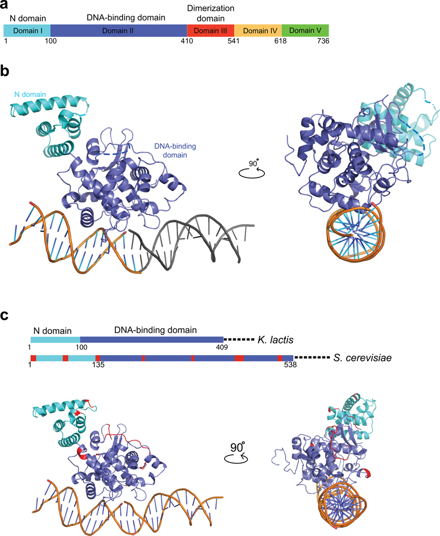
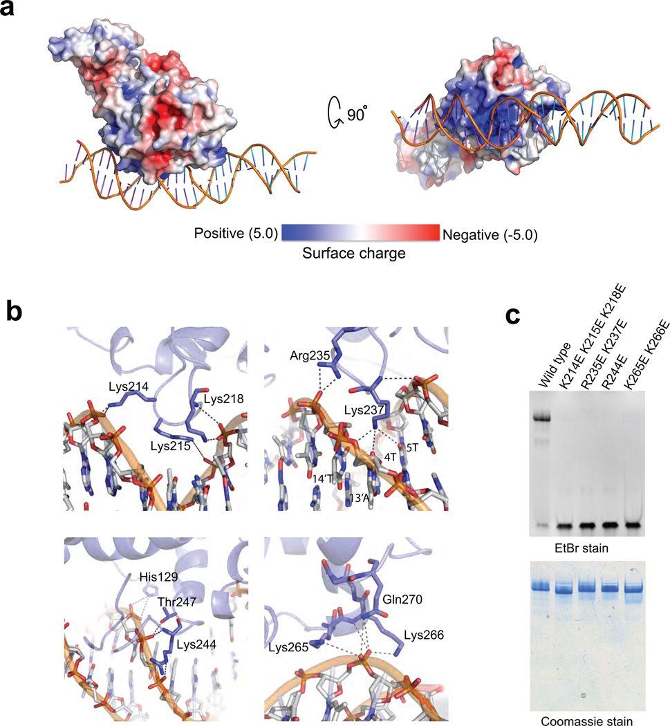
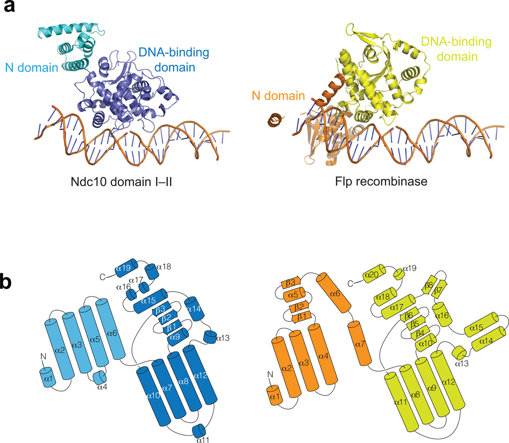
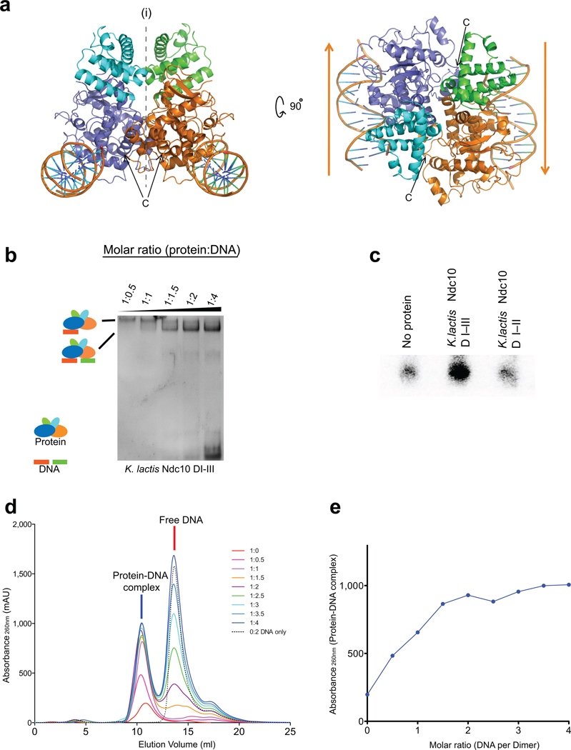
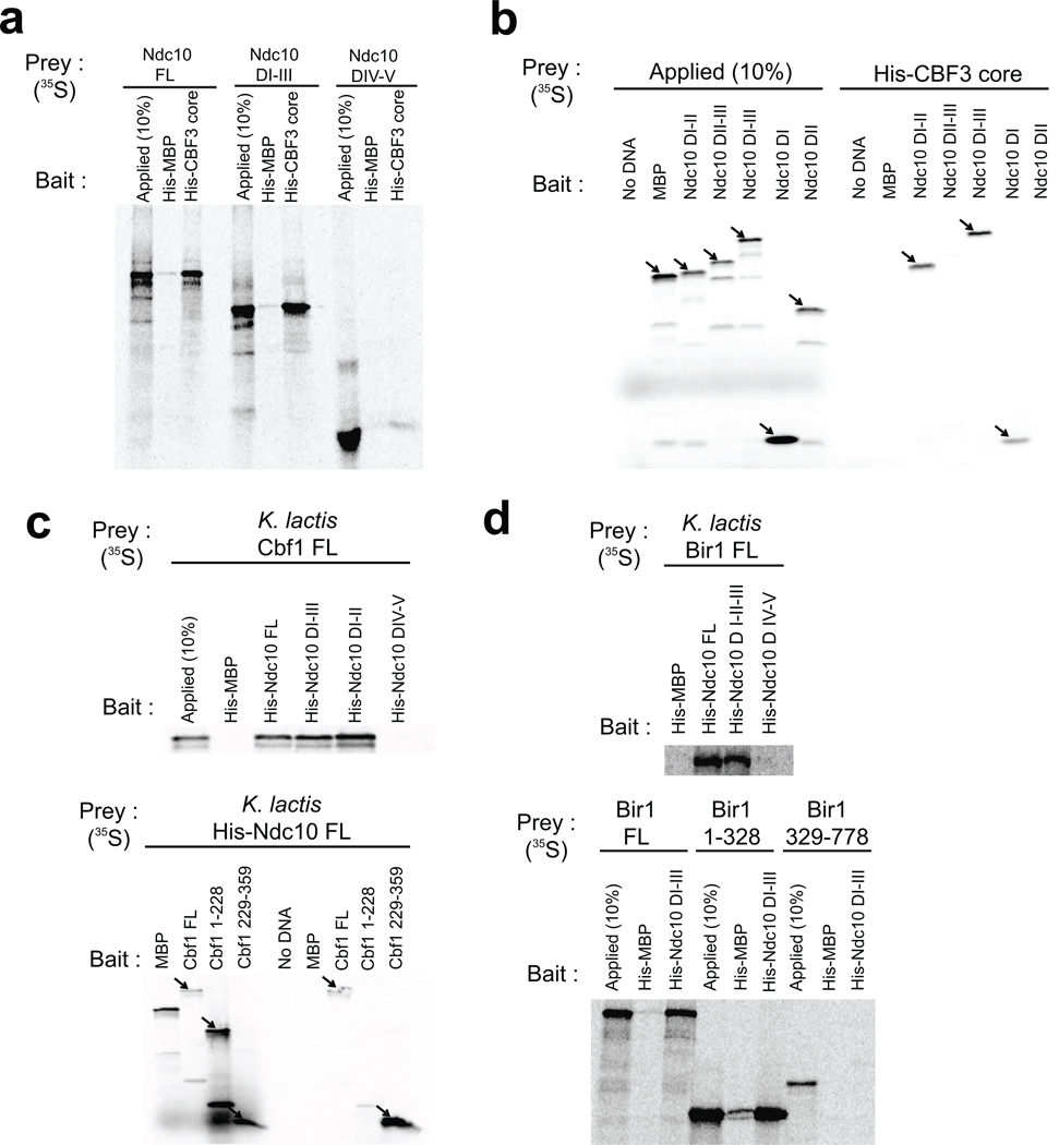
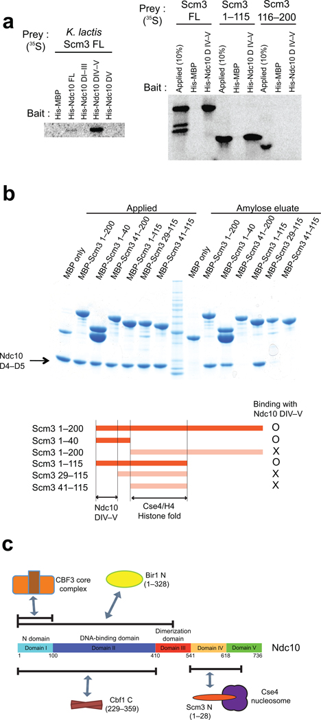
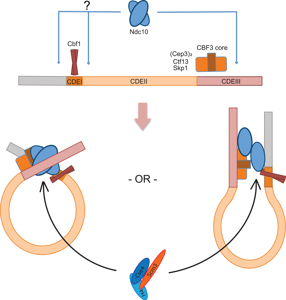
Kinetochores link centromeric DNA to spindle microtubules and ensure faithful chromosome segregation during mitosis. In point-centromere yeasts, the CBF3 complex Skp1-Ctf13-(Cep3)(2)-(Ndc10)(2) recognizes a conserved centromeric DNA element through contacts made by Cep3 and Ndc10. We describe here the five-domain organization of Kluyveromyces lactis Ndc10 and the structure at 2.8 Å resolution of domains I-II (residues 1-402) bound to DNA. The structure resembles tyrosine DNA recombinases, although it lacks both endonuclease and ligase activities. Structural and biochemical data demonstrate that each subunit of the Ndc10 dimer binds a separate fragment of DNA, suggesting that Ndc10 stabilizes a DNA loop at the centromere. We describe in vitro association experiments showing that specific domains of Ndc10 interact with each of the known inner-kinetochore proteins or protein complexes in budding yeast. We propose that Ndc10 provides a central platform for inner-kinetochore assembly.


Figures

Figure 1
Figure 1. Domains of K. lactis Ndc10 and crystal structure of DI–II
(a) Domain organization of Ndc10; numbers show residues at the domain boundaries derived either from limited proteolysis or from the crystal structure. (b) Structure of K. lactis Ndc10 (DI–II; 1–402) with 30 bp poly-dA:dT DNA. Domain I (N-domain, residues 1–100) is in cyan; domain II (DNA-binding domain, residues 101–402), in dark blue. Dashed lines represent disordered residues 36–39 and 283–292. A second, symmetry-related, 15 bp DNA fragment is shown in gray. The DNA has been modeled as polydA:dT (see text), with the sequence of 5’-TTAATTTATAAAATT-3’ (1–15) and 5’-AAATTTTATAAATTA-3’ (1’–15’), as indicated. (c) Sequence conservation of Ndc10 among point-centromere yeasts. Location of insertions (red) in S. cerevisiae Ndc10 DI–II with respect to K. lactis Ndc10 DI–II, shown both on a schematic representation of the primary sequence and on a ribbon representation of the structure. Illustration of all figures made with PYMOL (Delano Scientific, LLC).
Figure 2
Figure 2. Surface charge distribution and DNA contacts of Ndc10 DI–II
(a) Two views of the surface charge distribution of Ndc10 DI–II; bound DNA is shown in worm representation. (b) Sugar-phosphate backbone interactions. Residues involved in DNA contacts are labeled and shown in stick representation. (c) EMSA of wild-type and mutant Ndc10 (10% TBE acrylamide gel (w/v) stained sequentially with EtBr and Coomassie blue).
Figure 3
Figure 3. Structural alignment of K. lactis Ndc10 DI–II with Flp recombinases
(a) Monomer structure of Flp (PDB ID: 1M6X) aligned with the K. lactis Ndc10 DI–II. The N-domain and the DNA binding domain of Flp recombinase are colored in orange and yellow, respectively. In Flp, the DNA structure of the Holliday junction was replaced by 30 bp CDEIII DNA for simple comparison. (b) Folding diagrams of K. lactis Ndc10 DI–II and Flp recombinase. Secondary-structure elements are labeled according to their position in the polypeptide chain; domains colored as in panel a.
Figure 4
Figure 4. Dimerization of K. lactis Ndc10 DI–III
(a) Views of the likely Ndc10 DI–II dimer (symmetry axis along b in the C2221 space group). The subunits of the dimer contact different pseudocontinuous DNA duplexes. (b) EMSA of Ndc10 DI–III with increasing amounts of 30 bp CDEIII DNA. (c) DNA capture assay with two different labels. Either Ndc10 DI-III or Ndc10 DI-II was incubated with a mixture of equal amounts of biotinylated and unmodified CDEIII DNA, the including 32P-labeled product (10%). (d–e) Ratio of Ndc10 DI–III and CDEIII DNA determined by analytical size-exclusion chromatography.
Figure 5
Figure 5. Interactions of Ndc10-associated proteins or protein complexes in the inner kinetochore
(a–d) Ni2+ affinity pull-down of 35S-labeled, in vitro translated prey proteins with purified, His-tagged bait protein, analyzed by SDS-PAGE and visualized by phosphoimaging. Each panel includes a lane loaded with 10% of the in vitro translation reaction mixture (to monitor extent of synthesis) and either in vitro translated maltose-binding protein (MBP) as a prey or purified His-tagged MBP as a bait (negative controls).
Figure 6
Figure 6. Interaction of Ndc10 Domain IV–V with N-terminal Scm3
(a) Ni2+ affinity pull-down of 35S-labeled, in vitro translated Ndc10 proteins with purified, His-tagged Scm3 proteins, analyzed by SDS-PAGE and visualized by phosphoimaging. (b) In vitro amylose pull down of purified MBP tagged Scm3 proteins with Ndc10 domain IV–V. (c) Schematic overview of domain association of K. lactis Ndc10 with other kinetochore proteins. Ndc10 DI interacts with CBF3 core; Ndc10 DI–II, with Cbf1 (229–359) and Bir1p (1–328). Scm3 (1–28) associates with Ndc10 DIV–V but not with DV. Interaction of Cbf1 with Ndc10 was confirmed by analytical size-exclusion chromatography with purified proteins (Supplementary Fig. 6).
Figure 7
Figure 7. Schematic model of Ndc10 interactions on budding yeast centromeres
Cbf1 and CBF3 core recognize CDEI and CDEIII, respectively. Ndc10 does not have sequence-specific DNA contacts, but it binds in defined register through its interactions with Cbf1 and CBF3 core. We propose that these contacts bring CDEI and CDEIII together to form a loop. Two potential loop configurations are shown. The Scm3:Cse4:H4 heterotrimeric complex can be recruited through Scm3–Ndc10 interaction.

References

    1. Clarke L, Baum MP. Functional analysis of a centromere from fission yeast: a role for centromere-specific repeated DNA sequences. Mol. Cell Biol. 1990;10:1863–1872. - PMC - PubMed
    1. Fitzgerald-Hayes M, Clarke L, Carbon J. Nucleotide sequence comparisons and functional analysis of yeast centromere DNAs. Cell. 1982;29:235–244. - PubMed
    1. Murphy TD, Karpen GH. Localization of centromere function in a Drosophila minichromosome. Cell. 1995;82:599–609. - PMC - PubMed
    1. Willard HF. Centromeres of mammalian chromosomes. Trends Genet. 1990;6:410–416. - PubMed
    1. Meraldi P, McAinsh AD, Rheinbay E, Sorger PK. Phylogenetic and structural analysis of centromeric DNA and kinetochore proteins. Genome Biol. 2006;7:R23. - PMC - PubMed

Publication types

MeSH terms