. 2012 Sep 6;31(36):4054-66.
doi: 10.1038/onc.2011.563. Epub 2011 Dec 5.

EGFRvIII promotes glioma angiogenesis and growth through the NF-κB, interleukin-8 pathway

Affiliations

EGFRvIII promotes glioma angiogenesis and growth through the NF-κB, interleukin-8 pathway

R Bonavia et al. Oncogene. .

Abstract

Sustaining a high growth rate requires tumors to exploit resources in their microenvironment. One example of this is the extensive angiogenesis that is a typical feature of high-grade gliomas. Here, we show that expression of the constitutively active mutant epidermal growth factor receptor, ΔEGFR (EGFRvIII, EGFR*, de2-7EGFR) is associated with significantly higher expression levels of the pro-angiogenic factor interleukin (IL)-8 in human glioma specimens and glioma stem cells. Furthermore, the ectopic expression of ΔEGFR in different glioma cell lines caused up to 60-fold increases in the secretion of IL-8. Xenografts of these cells exhibit increased neovascularization, which is not elicited by cells overexpressing wild-type (wt)EGFR or ΔEGFR with an additional kinase domain mutation. Analysis of the regulation of IL-8 by site-directed mutagenesis of its promoter showed that ΔEGFR regulates its expression through the transcription factors nuclear factor (NF)-κB, activator protein 1 (AP-1) and CCAAT/enhancer binding protein (C/EBP). Glioma cells overexpressing ΔEGFR showed constitutive activation and DNA binding of NF-κB, overexpression of c-Jun and activation of its upstream kinase c-Jun N-terminal kinase (JNK) and overexpression of C/EBPβ. Selective pharmacological or genetic targeting of the NF-κB or AP-1 pathways efficiently blocked promoter activity and secretion of IL-8. Moreover, RNA interference-mediated knock-down of either IL-8 or the NF-κB subunit p65, in ΔEGFR-expressing cells attenuated their ability to form tumors and to induce angiogenesis when injected subcutaneously into nude mice. On the contrary, the overexpression of IL-8 in glioma cells lacking ΔEGFR potently enhanced their tumorigenicity and produced highly vascularized tumors, suggesting the importance of this cytokine and its transcription regulators in promoting glioma angiogenesis and tumor growth.


Conflict of interest statement

Conflict of interest

The authors declare no conflict of interest.

Figures
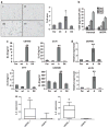
Figure 1
Figure 1
ΔEGFR upregulates IL-8 and induces angiogenesis in glioma cells. (a) IHC analysis by CD31 staining of intracranial xenografts: representative fields of U87Par (I), U87wt (II), U87Δ (III) or U87DK (IV) xenografts (left) and relative quantification of vessel density expressed as percentage of stained area. (b) Quantification by ELISA of VEGF secretion in U87Par and U87Δ after 1, 2, 3 and 4 days. (c) IL-8 concentration in CM generated from U87MG, U178, U373 and LNZ308 GBM cell lines engineered to overexpress wtEGFR (wt), ΔEGFR (Δ) or a dead kinase version of ΔEGFR was determined by ELISA. (d) IL-8 expression in U87MG and U178 cell lines and derivatives was determined by real-time PCR. (e) Comparison of IL-8 relative expression in ΔEGFR-positive and -negative GBM clinical samples (left) and GBM tumorspheres (right) determined by real-time PCR. *P<0.05; **P<0.01.
Figure 2
Figure 2
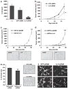
ΔEGFR promotes glioma angiogenesis and tumor growth through induction of IL-8 expression. (a) ELISA quantification of IL-8 secretion in U87Δ cells after transfecting with GFP or IL-8 siRNAs. Cells not transfected were included as negative control (−). (b) Tumor growth curve after subcutaneous injection of U87Δ cells transfected with 25 nM of GFP or IL-8 siRNAs. (c) Top: tumor growth curve after subcutaneous injection of U87wt cells infected with empty vector (pBABE) or engineered to overexpress IL-8 (IL-8). Bottom: representative fields of CD31 immunohistochemistry on sections of U87wt-pBABE (left) or U87wt-IL-8 (right) xenografts. (d) Top: tumor growth curve after subcutaneous injection of U87MG cells infected with empty vector (pBABE) or engineered to overexpress IL-8 (IL-8). Bottom: CD31 immunohistochemistry on corresponding xenografts, U87Par-pBABE (left) and U87Par-IL-8 (right). (e) Quantification of vessel relative area (left) and CD31 immunohistochemistry (right) of xenografts obtained in (b). (f) Representative images of HUVEC cells treated with CM from U87Δ cells transfected with GFP or IL-8 siRNA showing in vitro tube formation. *P<0.05; **P<0.01.
Figure 3
Figure 3
ΔEGFR activates regulatory elements in IL-8 promoter. (a) Scheme of IL-8 proximal promoter (from −400 bp) showing the responsive elements that were mutated in the following experiments. (b) U87Par (Parental) and U87Δ (Δ) cells were transfected with a reporter construct consisting of the luciferase gene under the control of wild-type IL-8 promoter (WT) or the IL-8 promoter mutated at binding sites for individual transcription factors indicated; transcriptional activity was measured by luciferase assay of cellular extracts after 24 h of serum starvation. Data represent mean±s.e.m. Luciferase activity assay is expressed in relative light units (RLU) normalized to corresponding readings in U87Par. **P<0.01.
Figure 4
Figure 4
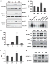
NF-κB, AP-1 and C/EBP are activated in ΔEGFR-expressing glioma cells. (a) Phosphorylation of Ser 536 of p65 (ph-p65), indicative of NF-κB activation, was analyzed by western blot in U87MG cell line derivatives grown in media supplemented with 10% FBS or serum-starved for 24 h or 48 h. (b) U87MG cells and their derivatives were transfected with the NF-κB reporter and transcriptional activity was measured by luciferase activity assay. (c) Activation of NF-κB in U87MG cells and their derivatives serum-starved for 48 h and then treated where indicated with 50 ng/ml TNF-α or 50 ng/ml EGF for 15 min. Activation of NF-κB was analyzed by binding of nuclear extracts to a radiolabeled NF-κB consensus oligonucleotide. (d) Promoter assay using U87MG cells and their derivatives transfected with an AP-1 reporter plasmid followed by quantification of luciferase activity. (e) Phosphorylation of JNK (ph-JNK) and c-Jun expression were analyzed by western blot in U87MG cell line derivatives, showing increased JNK activation and high levels of c-Jun in ΔEGFR-expressing cells. (f) Promoter assay using U87MG cells and their derivatives transfected with a C/EBP reporter plasmid followed by quantification of luciferase activity. (g) Analysis of C/EBPβ expression by western blot analysis in U87MG cell line derivatives. Data represent mean±s.e.m. Luciferase activity is expressed in RLU after normalizing by Renilla luciferase reporter activity. *P<0.05; **P<0.01.
Figure 5
Figure 5
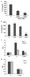
Inhibition of NF-κB and JNK/cJun reduces IL-8 promoter activity and secretion in ΔEGFR-expressing glioma cells. (a) Quantification of IL-8 secretion by ELISA in supernatants of U87Δ cells serum-starved for 48 h and treated for 24 h with 10 or 20 μM of BAY11-7082, a pharmacological NF-κB inhibitor. Cells not treated (−) were used as control. (b) IL-8 quantification by ELISA in supernatants of U87Δ (Δ) cells treated with inhibitors of the different MAP kinases: PD=PD98059, MEK/ERK inhibitor (10 μM); SB=SB203580, p38 inhibitor (10 μM); SP=SP600125, JNK inhibitor (10 μM). (c) IL-8 promoter reporter and a NF-κB reporter construct (pNFκB-Luc, Clontech, containing multiple copies of the NF-κB consensus sequence) were transfected into U87Par (Par), U87Δ cells stably infected with empty pCLBabePuro (Δ-pCLBP) or U87Δ stably infected with pCLBabe-Puro containing IκB super-repressor (Δ-IκBsr) and transcriptional activity was measured by luciferase assay. (d) IL-8 promoter reporter or AP-1 reporter (pAP1-Luc, Clontech) were co-transfected into U87Δ with c-Jun dominant-negative (cJundn) or the same amount of empty vector (−) and transcriptional activity was measured by luciferase activity assay. Data represent mean±s.e.m. Luciferase activity assays are expressed in RLU. *P<0.05; **P<0.01.
Figure 6
Figure 6
Ras and PI-3K involvement in the activation of IL-8 promoter. (a) Transcriptional activity of IL-8 promoter in U87Δ cells infected with different retroviruses: pBabe-Puro empty vector (pBP) or pBabe-Puro-expressing p85ΔSH2, Ras17N or IκBsr. (b) Western blot analysis of Akt phosphorylation from lysates of U87Δ cells infected with pBabe-Puro empty vector (pBP) or containing p85ΔSH2, and Ras17N. (c) IL-8 quantification by ELISA in supernatants of U87Par (Par) and U87Δ (Δ) cells serum-starved for 24 h, replenished with fresh serum-free media and vehicle treated (dimethylsulphoxide (DMSO)) or treated with LY294002 10 μM (LY) for 4 h or 24 h. (d) Western blot analysis of Akt phosphorylation of lysates fromU87Δ treated for different times with LY294002 10 μM. (e) Transcriptional activity of IL-8 promoter, and AP-1 and NF-κB reporter constructs in U87Δ cells infected with pBabe-Puro empty vector (pBP) or pBabe-Puro-expressing Ras17N. (f) Transcriptional activity of AP-1, NF-κB and C/EBP reporter constructs in U87Δ cells treated 24 h with vehicle (DMSO) or the JNK inhibitor SP600125 (10 μM). Data represent mean±s.e.m. Luciferase activity is expressed in relative light units (RLU). *P<0.05; **P<0.01.
Figure 7
Figure 7
NF-κB activation is important for ΔEGFR-mediated tumorigenesis and angiogenesis. (a) Western blot analysis of p65 expression in U87Δ cells transfected with a GFP shRNA or shRNAs against the NF-κB p65 subunit. (b) Top: tumor growth curve after subcutaneous injection of U87Δ cells stably expressing a shRNA against GFP or with a shRNA against NF-κB p65 (shp65). Bottom: CD31 immunohistochemistry on corresponding xenografts, U87Δ-shGFP (right) and U87Δ-shp65 (left). (c) ELISA quantification ofIL-8 in tumor lysates generated in panel (b). Data represent mean±s.e.m. **P<0.01.

Similar articles

Cited by

References

    1. Antonyak MA, Moscatello DK, Wong AJ. Constitutive activation of c-Jun N-terminal kinase by a mutant epidermal growth factor receptor. J Biol Chem. 1998;273:2817–2822. - PubMed
    1. Bachoo RM, Maher EA, Ligon KL, Sharpless NE, Chan SS, You MJ, et al. Epidermal growth factor receptor and Ink4a/Arf: convergent mechanisms governing terminal differentiation and transformation along the neural stem cell to astrocyte axis. Cancer Cell. 2002;1:269–277. - PubMed
    1. Bancroft CC, Chen Z, Yeh J, Sunwoo JB, Yeh NT, Jackson S, et al. Effects of pharmacologic antagonists of epidermal growth factor receptor, PI3K and MEK signal kinases on NF-kappaB and AP-1 activation and IL-8 and VEGF expression in human head and neck squamous cell carcinoma lines. Int J Cancer. 2002;99:538–548. - PubMed
    1. Biernat W, Huang H, Yokoo H, Kleihues P, Ohgaki H. Predominant expression of mutant EGFR (EGFRvIII) is rare in primary glioblastomas. Brain Pathol. 2004;14:131–136. - PMC - PubMed
    1. Biswas DK, Cruz AP, Gansberger E, Pardee AB. Epidermal growth factor-induced nuclear factor kappa B activation: a major pathway of cell-cycle progression in estrogen-receptor negative breast cancer cells. Proc Natl Acad Sci USA. 2000;97:8542–8547. - PMC - PubMed

Publication types

MeSH terms