. 2012 Aug 9;31(32):3679-95.
doi: 10.1038/onc.2011.545. Epub 2011 Dec 5.

Suppression of Tumorigenicity-14, encoding matriptase, is a critical suppressor of colitis and colitis-associated colon carcinogenesis

Affiliations

Suppression of Tumorigenicity-14, encoding matriptase, is a critical suppressor of colitis and colitis-associated colon carcinogenesis

P Kosa et al. Oncogene. .

Abstract

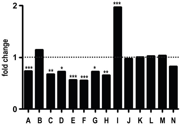
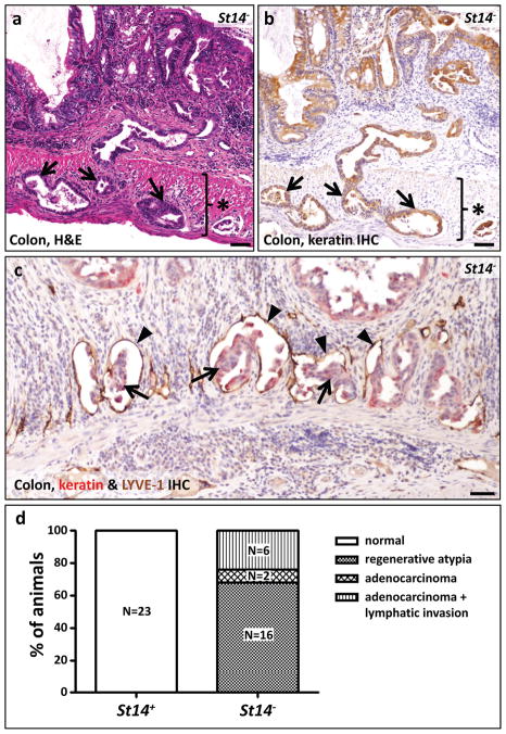
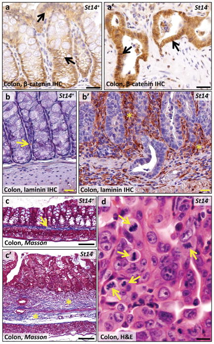
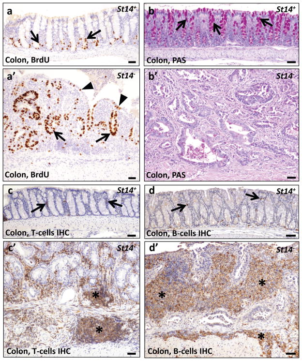
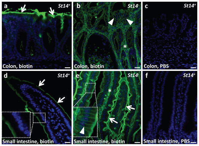
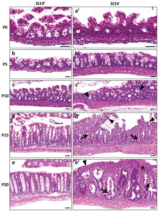
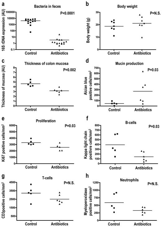
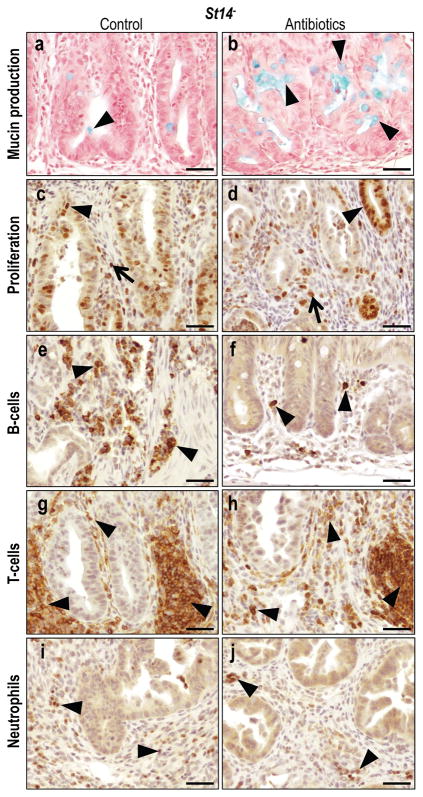
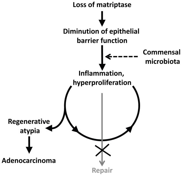
Colitis-associated colorectal cancers are an etiologically distinct subgroup of colon cancers that occur in individuals suffering from inflammatory bowel disease and arise as a consequence of persistent exposure of hyperproliferative epithelial stem cells to an inflammatory microenvironment. An intrinsic defect in the intestinal epithelial barrier has been proposed to be one of several factors that contribute to the inappropriate immune response to the commensal microbiota that underlies inflammatory bowel disease. Matriptase is a membrane-anchored serine protease encoded by Suppression of Tumorigenicity-14 (ST14) that strengthens the intestinal epithelial barrier by promoting tight junction formation. Here, we show that intestinal epithelial-specific ablation of St14 in mice causes formation of colon adenocarcinoma with very early onset and high penetrance. Neoplastic progression is preceded by a chronic inflammation of the colon that resembles human inflammatory bowel disease and is promoted by the commensal microbiota. This study demonstrates that inflammation-associated colon carcinogenesis can be initiated and promoted solely by an intrinsic intestinal permeability barrier perturbation, establishes St14 as a critical tumor-suppressor gene in the mouse gastrointestinal tract and adds matriptase to the expanding list of pericellular proteases with tumor-suppressive functions.


Conflict of interest statement

Conflict of interest

The authors declare no competing financial interests in relation to the work described.

Figures

Figure 1
Figure 1. Matriptase expression is downregulated in human colon adenomas and adenocarcinomas
Expression of ST14, encoding matriptase, in 14 gene expression array studies of human colon adenomas and adenocarcinomas. Data are expressed as fold change relative to corresponding normal tissue. *P<0.05, **P<0.01, ***P<0.001. See Supplementary Table 1 for details and references.
Figure 2
Figure 2. Rapid and spontaneous malignant transformation of St14-ablated colonic epithelium
(a) Representative example of adenocarcinoma in the large intestine of an eight week old St14 mouse. Tumor cells invading the muscularis externa (star) are shown with arrows. (b) The epithelial origin of the tumor cells invading the muscularis externa (star) is demonstrated by immunohistochemical staining for keratin (examples with arrows). (c) Combined immunohistochemical staining for keratin in red (examples with arrows) and the lymphatic vessel marker LYVE-1 in brown (examples with arrowheads) shows invasion of malignant cells into lymphatic vessels of a seven week old St14 mouse. Scale bar for a, b, and c = 100 μm. (d) Enumeration of colonic lesions in four to 18 week old St14+ (left) and littermate St14 mice (right), showing adenocarcinoma with lymphatic invasion in 6, adenocarcinoma without lymphatic invasion in 2, and regenerative atypia in the remaining 16 St14 mice. See Table 1 for additional details.
Figure 3
Figure 3. Characterization of matriptase ablation-associated colon adenocarcinoma
(a,a′) Immunohistochemical staining of eight week old St14+ (a) and littermate St14 (a′) colons for β-catenin shows a membrane-associated β-catenin localization in St14+ epithelial cells (arrows in a), as compared to cytoplasmic and nuclear localization in adenocarcinomas of St14 colons (examples with arrows in a′). (b,b′) Immunohistochemical staining for the basement membrane marker laminin in 15 week old St14+ (b) and littermate St14 (b′) mice shows the normal appearance of the basement membrane (example with arrow in b) in St14+ mice. Loss of matriptase expression leads to increased deposition of laminin (examples with stars in b′) and loss of normal structure of the basement membrane. (c,c′) Masson Trichrome staining of the colon of six week old St14+ (c) and littermate St14 (c′) mice shows connective tissue in the submucosa of a normal colon (example with arrow in c) and fibrosis of both the mucosa and submucosa of St14 colon (examples with stars in c′). (d) High magnification shows the cytological appearance of adenocarcinomas of St14 mice. Atypical mitosis is shown by arrows. Scale bar = 200 μm (a, a′,b,b′,c,c′) and 20 μm (d).
Figure 4
Figure 4. Characterization of matriptase ablation-associated colon adenocarcinoma
(a,a′) BrdU staining of eight week old St14+ (a) and littermate St14 (a′) mice shows proliferation restricted to the bottom of the crypts of normal colons (examples with arrows in a). In St14 colon, proliferating cells are found both in the bottom (examples with arrows in a′) and distal parts of crypts (examples with arrowheads in a′). (b,b) Periodic Acid-Schiff (PAS) staining of mucopolysaccharides produced by differentiated goblet cells in the colon of eleven week old St14+ (b) and littermate St14 (b′) mice. Red staining shows mucin in the normal colon (arrows in b). Absence of red staining in (b′) indicates cessation of mucin production in matriptase-ablated colon. (c,c′,d,d′) Immunohistochemical staining for T-cells (c,c′) and B-cells (d,d′) in, respectively, seven and 15 week old St14+ (c, d) and littermate St14 (c′,d′) colons. Baseline levels of T-and B-cells in the lamina propria of St14+ colon (examples with arrows in d and c) and abundance of T- and B-cells in both mucosa and submucosa of St14 colons (examples with stars in c′,d′). Scale bar = 100 μm.
Figure 5
Figure 5. Matriptase-ablated colon is leaky
The lumen of the colon and small intestine of weaning age St14+ and littermate St14animals was injected with Sulfo-NHS-LC-Biotin in PBS (a,b,d,e) or PBS (c,f). After three min, the intestine was excised, sectioned, and stained for biotin (green). Nuclei were stained with 4,6-diamino-2-phenylindol (blue). Arrows in a, d, and e show biotin bound to the surface of the mucosa. Arrowheads in b and the inset in e show biotin labeling of the basolateral membrane of polarized epithelial cells. The diffusion of biotin into intercellular space was not observed in the normal colon or small intestine (a,d, also compare insets in d and e). Stars show biotin labeling of connective tissue of both matriptase-ablated colon (b) and small intestine (e). There was no signal for biotin in colon and small intestine (c,f) injected with PBS. Scale bar = 20 μm.
Figure 6
Figure 6. Progressive postnatal loss of epithelial integrity of matriptase-ablated colon precedes malignant transformation
Histological appearance of St14+ (ae) and littermate St14 (a′-e′) colons at postnatal day 0 (a,a′), 5 (b,b′), 10 (c,c′), 15 (d,d′), and 20 (e,e′). No histological differences can be observed between normal and matriptase-ablated colon at days 0 and 5 (compare a and a′, b and b′). At day 10, St14 colons show sporadic foci of detaching and apoptotic cells (arrowheads in c′). This phenotype is significantly stronger at days 15 and 20 with extensive anoikis (arrowheads in d′), apoptotic cells (arrows in d′, e′), ulcerations (arrowhead in e′) and inflammatory cell infiltrates (star in e′). Scale bar = 100 μm.
Figure 7
Figure 7. The resident microbiota contributes to preneoplastic progression of matriptase-ablated colon
Littermate St14 mice were kept on regular water (control in ah) or treated with a combination of ampicillin, neomycin, metronidazole, and vancomycin in the drinking water (antibiotics in ah) for two weeks starting immediately after weaning. The animals were euthanized, the feces was used for the isolation of bacterial DNA, and the colonic tissue was subjected to quantitative histomorphometric analysis. (a) PCR quantification of 16S bacterial ribosomal DNA shows a 1 500-fold decrease in the intestinal microbiota of antibiotics treated (N=15) compared to control (N=13) mice. (b) Body weight of antibiotics treated (N=7) and control (N=7) is similar. (c) Decreased thickness of the mucosa of antibiotics treated (N=6) compared to control (N=5) mice. (df) Preservation of mucin production (d), decreased proliferation (e), and decreased infiltration of B-cells (f), T-cells (g) and neutrophils (h) in antibiotics treated (N=6) compared to (N=5) mice. Statistical significance was calculated by Student’s t-test (two-tailed) (ac, eh), and non-parametric Mann-Whitney U-test (two-tailed) (d), N.S. = not significant.
Figure 8
Figure 8. Histological appearance of antibiotics treated matriptase-ablated colon
(a,b) Alcian Blue staining of mucin produced by differentiated goblet cells in untreated (a) and antibiotics treated (b) St14 colons. Arrowheads point to mucin (blue). (c,d) Immunohistochemical staining for Ki67 in untreated (c) and antibiotics treated (d) St14mice show significantly decreased rates of proliferation of both epithelial cells (arrowheads in c,d) and connective tissue cells (arrows in c,d). (ej) Immunohistochemical staining for B-cells (e,f), T-cells (g,h), and neutrophils (i,j) in untreated (e,g,i) and antibiotics treated (f,h,j) St14 colons shows reduced chronic (examples with arrowheads in eh) and acute (examples with arrowheads in i and j) inflammatory cell infiltration. Scale bar = 50 μm.
Figure 9
Figure 9. Model for matriptase ablation-induced colon carcinogenesis
Loss of matriptase from intestinal epithelium compromises epithelial barrier function thereby causing exposure of the commensal microbiota to resident immune cells. This triggers a repair response that includes activation of local inflammatory circuits and colonic stem cell activation. This response is perpetual, rather than transient, due to the intrinsic inability of matriptase-ablated to form a functional barrier. Persistent hyperproliferation of colonic stem cells within a DNA damaging chronic inflammatory microenvironment causes the formation of adenocarcinoma.

Similar articles

Cited by

References

    1. Acuff HB, Sinnamon M, Fingleton B, Boone B, Levy SE, Chen X, et al. Analysis of host- and tumor-derived proteinases using a custom dual species microarray reveals a protective role for stromal matrix metalloproteinase-12 in non-small cell lung cancer. Cancer Res. 2006;66:7968–7975. - PubMed
    1. Andreasen PA, Egelund R, Petersen HH. The plasminogen activation system in tumor growth, invasion, and metastasis. Cell Mol Life Sci. 2000;57:25–40. - PMC - PubMed
    1. Balbin M, Fueyo A, Tester AM, Pendas AM, Pitiot AS, Astudillo A, et al. Loss of collagenase-2 confers increased skin tumor susceptibility to male mice. Nat Genet. 2003;35:252–257. - PubMed
    1. Berg DJ, Davidson N, Kuhn R, Muller W, Menon S, Holland G, et al. Enterocolitis and colon cancer in interleukin-10-deficient mice are associated with aberrant cytokine production and CD4(+) TH1-like responses. J Clin Invest. 1996;98:1010–1020. - PMC - PubMed
    1. Bhatt AS, Welm A, Farady CJ, Vasquez M, Wilson K, Craik CS. Coordinate expression and functional profiling identify an extracellular proteolytic signaling pathway. Proc Natl Acad Sci U S A 2007 - PMC - PubMed

Publication types

MeSH terms