

Quercetin-3-methyl ether suppresses proliferation of mouse epidermal JB6 P+ cells by targeting ERKs
- PMID: 22139441
- PMCID: PMC3271267
- DOI: 10.1093/carcin/bgr281
Quercetin-3-methyl ether suppresses proliferation of mouse epidermal JB6 P+ cells by targeting ERKs
Abstract
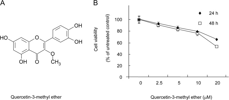
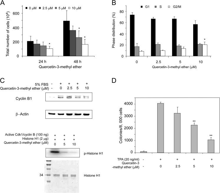
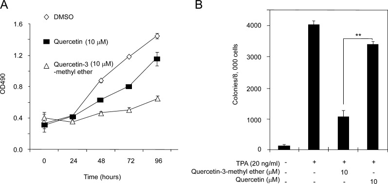
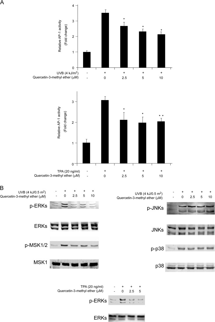
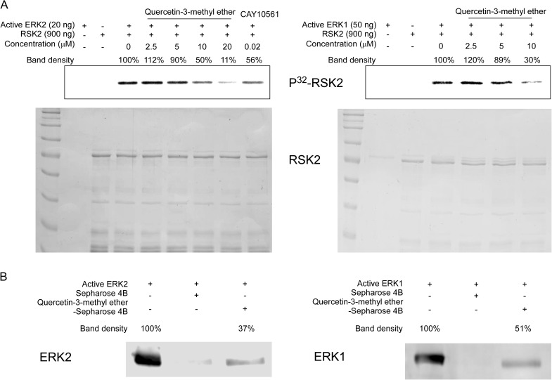
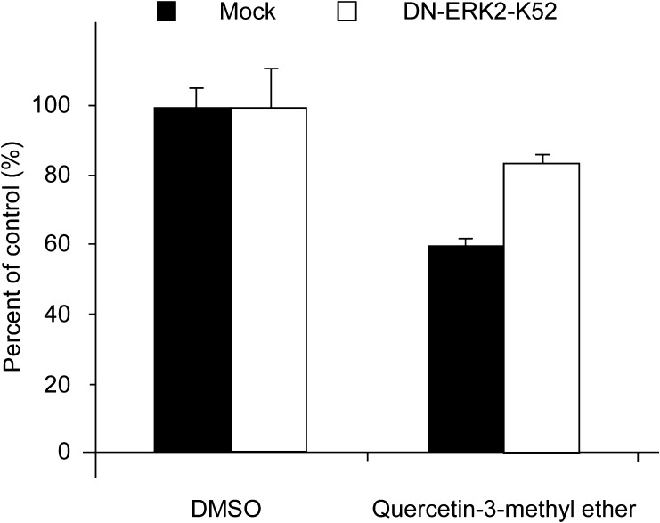
Chemoprevention has been acknowledged as an important and practical strategy for the management of skin cancer. Quercetin-3-methyl ether, a naturally occurring compound present in various plants, has potent anticancer-promoting activity. We identified this compound by in silico virtual screening of the Traditional Chinese Medicine Database using extracellular signal-regulated kinase 2 (ERK2) as the target protein. Here, we showed that quercetin-3-methyl ether inhibited proliferation of mouse skin epidermal JB6 P+ cells in a dose- and time-dependent manner by inducing cell cycle G(2)-M phase accumulation. It also suppressed 12-O-tetradecanoylphorbol-13-acetate-induced neoplastic cell transformation in a dose-dependent manner. Its inhibitory effect was greater than quercetin. The activation of activator protein-1 was dose-dependently suppressed by quercetin-3-methyl ether treatment. Western blot and kinase assay data revealed that quercetin-3-methyl ether inhibited ERKs kinase activity and attenuated phosphorylation of ERKs. Pull-down assays revealed that quercetin-3-methyl ether directly binds with ERKs. Furthermore, a loss-of-function ERK2 mutation inhibited the effectiveness of the quercetin-3-methyl ether. Overall, these results indicated that quercetin-3-methyl ether exerts potent chemopreventive activity by targeting ERKs.
Figures
Similar articles
-
Raf and MEK protein kinases are direct molecular targets for the chemopreventive effect of quercetin, a major flavonol in red wine.Cancer Res. 2008 Feb 1;68(3):946-55. doi: 10.1158/0008-5472.CAN-07-3140. Cancer Res. 2008. PMID: 18245498 Free PMC article.
-
The extracellular-signal-regulated protein kinases (Erks) are required for UV-induced AP-1 activation in JB6 cells.Oncogene. 1999 May 6;18(18):2828-35. doi: 10.1038/sj.onc.1202639. Oncogene. 1999. PMID: 10362253
-
Quercetin-3-methyl ether inhibits esophageal carcinogenesis by targeting the AKT/mTOR/p70S6K and MAPK pathways.Mol Carcinog. 2018 Nov;57(11):1540-1552. doi: 10.1002/mc.22876. Epub 2018 Aug 28. Mol Carcinog. 2018. PMID: 30035335
-
Activator protein 1 (AP-1)- and nuclear factor kappaB (NF-kappaB)-dependent transcriptional events in carcinogenesis.Free Radic Biol Med. 2000 May 1;28(9):1338-48. doi: 10.1016/s0891-5849(00)00220-3. Free Radic Biol Med. 2000. PMID: 10924853 Review.
-
PI-3 kinase in signal transduction, cell transformation, and as a target for chemoprevention of cancer.Anticancer Res. 1999 Sep-Oct;19(5A):3743-7. Anticancer Res. 1999. PMID: 10625951 Review.
Cited by
-
Signal transduction and molecular targets of selected flavonoids.Antioxid Redox Signal. 2013 Jul 10;19(2):163-80. doi: 10.1089/ars.2013.5251. Epub 2013 Apr 15. Antioxid Redox Signal. 2013. PMID: 23458437 Free PMC article. Review.
-
Inhibition of Breast Cancer Cell Proliferation and In Vitro Tumorigenesis by a New Red Apple Cultivar.PLoS One. 2015 Aug 18;10(8):e0135840. doi: 10.1371/journal.pone.0135840. eCollection 2015. PLoS One. 2015. PMID: 26284516 Free PMC article.
-
Neuroprotective Assessment of Moringa oleifera Leaves Extract against Oxidative-Stress-Induced Cytotoxicity in SHSY5Y Neuroblastoma Cells.Plants (Basel). 2021 Apr 28;10(5):889. doi: 10.3390/plants10050889. Plants (Basel). 2021. PMID: 33925070 Free PMC article.
-
Influence of Harvesting Stage on Phytochemical Composition, Antioxidant, and Antidiabetic Activity of Immature Ceratonia siliqua L. Pulp from Béni Mellal-Khénifra Region, Morocco: In Silico, In Vitro, and In Vivo Approaches.Curr Issues Mol Biol. 2024 Sep 29;46(10):10991-11020. doi: 10.3390/cimb46100653. Curr Issues Mol Biol. 2024. PMID: 39451533 Free PMC article.
-
Molecular docking analysis of known flavonoids as duel COX-2 inhibitors in the context of cancer.Bioinformation. 2015 Dec 31;11(12):543-9. doi: 10.6026/97320630011543. eCollection 2015. Bioinformation. 2015. PMID: 26770028 Free PMC article.
References
-
- Jung SK, et al. Myricetin suppresses UVB-induced skin cancer by targeting Fyn. Cancer Res. 2008;68:6021–6029. - PubMed
-
- Assefa Z, et al. Differential stimulation of ERK and JNK activities by ultraviolet B irradiation and epidermal growth factor in human keratinocytes. J. Invest. Dermatol. 1997;108:886–891. - PubMed
-
- Zhang W, et al. MAPK signal pathways in the regulation of cell proliferation in mammalian cells. Cell Res. 2002;12:9–18. - PubMed
-
- Sebolt-Leopold JS, et al. Targeting the mitogen-activated protein kinase cascade to treat cancer. Nat. Rev. Cancer. 2004;4:937–947. - PubMed
Publication types
MeSH terms
Substances
Grants and funding
LinkOut - more resources
Full Text Sources
Research Materials
Miscellaneous
