. 2012 Nov;22(11):2662-71.
doi: 10.1093/cercor/bhr344. Epub 2011 Dec 1.

Contralesional hemisphere control of the proximal paretic upper limb following stroke

Affiliations

Contralesional hemisphere control of the proximal paretic upper limb following stroke

Lynley V Bradnam et al. Cereb Cortex. 2012 Nov.

Abstract

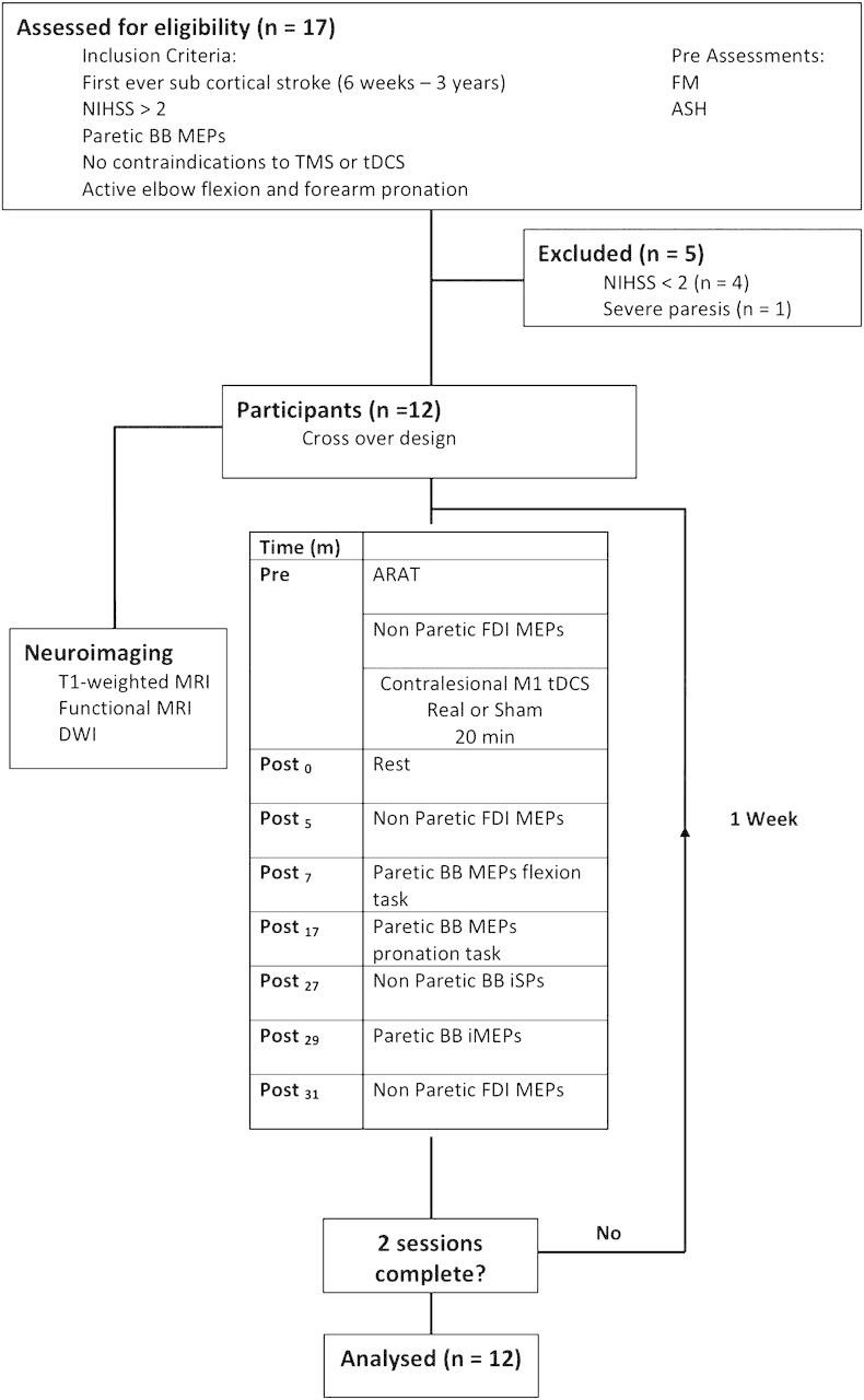
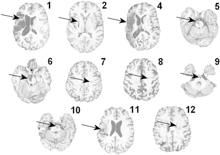
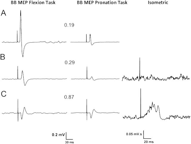
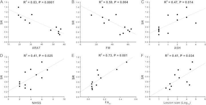
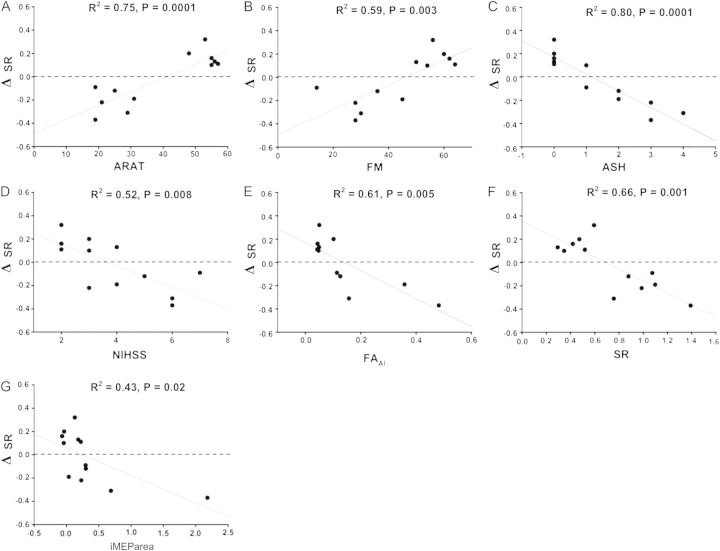
Cathodal transcranial direct current stimulation (c-tDCS) can reduce excitability of neurons in primary motor cortex (M1) and may facilitate motor recovery after stroke. However, little is known about the neurophysiological effects of tDCS on proximal upper limb function. We hypothesized that suppression of contralesional M1 (cM1) excitability would produce neurophysiological effects that depended on the severity of upper limb impairment. Twelve patients with varying upper limb impairment after subcortical stroke were assessed on clinical scales of upper limb spasticity, impairment, and function. Magnetic resonance imaging was used to determine lesion size and fractional anisotropy (FA) within the posterior limbs of the internal capsules indicative of corticospinal tract integrity. Excitability within paretic M1 biceps brachii representation was determined from motor-evoked potentials during selective isometric tasks, after cM1 sham stimulation and after c-tDCS. These neurophysiological data indicate that c-tDCS improved selective proximal upper limb control for mildly impaired patients and worsened it for moderate to severely impaired patients. The direction of the neurophysiological after effects of c-tDCS was strongly related to upper limb spasticity, impairment, function, and FA asymmetry between the posterior limbs of the internal capsules. These results indicate systematic variation of cM1 for proximal upper limb control after stroke and that suppression of cM1 excitability is not a "one size fits all" approach.


Figures

Figure 1.
Figure 1.
The experimental protocol for patients. Pre, post = before and after tDCS.
Figure 2.
Figure 2.
Structural T1-weighted images in the axial plane are shown at the level of the lesion for each patient. Lesions are indicated by the arrows. Patient numbers correspond with Table 1. Note there is no T1-weighted image for patient 3.
Figure 3.
Figure 3.
Averaged paretic or nondominant BB MEPs from representative participants showing BB MEPs from the flexion and (left column), pronation tasks (middle column), and rectified BB iMEPs from the isometric task in patients (right column). Contralateral MEP traces are shown from (A) a healthy participant (#3, Table 2), (B) a mildly impaired patient (#9, Tables 1 and 3), and (C) a moderately impaired patient (#4). Ipsilateral (iMEP) traces are shown from a mildly impaired patient (#9) and a severely impaired patient (#1). The SR value is indicated between the BB MEP traces. Note the different calibration bars for MEPs and iMEPs.
Figure 4.
Figure 4.
Regressions with SR following sham tDCS. (A) SR and ARAT upper limb function score. Larger SRs were associated with lower ARAT scores, indicating lower paretic arm function. (B) SR and FM upper limb impairment score. Larger SRs were associated with low FM scores and greater upper limb impairment. (C) SR and ASH. Larger SRs were associated with greater spasticity in paretic BB. (D) SR and NIHSS. Larger SRs were associated with higher NIHSS scores, indicating greater upper limb impairment. (E) SR and FAAI. Larger SRs were associated with higher FAAI, indicating reduced corticospinal tract integrity from the ipsilesional hemisphere. (F) SR and lesion volume (log transformed). Larger SRs were associated with larger stroke lesions.
Figure 5.
Figure 5.
Regressions with ΔSR. Positive ΔSR indicates improved selective muscle activation after contralesional c-tDCS and negative ΔSR indicates a worsening of selective muscle activation after contralesional c-tDCS. The dashed line represents no change, ΔSR = 0. (A) ΔSR and ARAT. SR improved for those with mildly impaired upper limb function worsened for those with ARAT scores < 40. (B) ΔSR and FM score. SR tended to improve for patients with mild upper limb impairment and worsen for those with FM < 50. (C) ΔSR and ASH. SR tended to improve for patients without elbow flexor spasticity and worsen for those with ASH > 1. (D) ΔSR and NIHSS. SR tended to worsen for patients with NIHSS of 5 or more. (E) ΔSR and FAAI. SR tended to improve for patients with good ipsilesional corticospinal tract integrity as indicated by FAAI < 0.11 but worsen for those with reduced ipsilesional corticospinal tract integrity. (F) ΔSR and SR. SR tended to improve in those with good initial selective muscle activation and worsen for those with poor initial selective muscle activation. (G) ΔSR and iMEParea. SR tended to improve for patients with small iMEPs in the paretic BB and worsen for those with large iMEPs. All other abbreviations as in text.

References

    1. Ackerley SJ, Stinear CM, Barber PA, Byblow WD. Combining theta burst stimulation with training after subcortical stroke. Stroke. 2010;41:1568–1572. - PubMed
    1. Alagona G, Delvaux V, Gerard P, De Pasqua V, Pennisi G, Delwaide PJ, Nicoletti F, Maertens de Noordhout A. Ipsilateral motor responses to focal transcranial magnetic stimulation in healthy subjects and acute-stroke patients. Stroke. 2001;32:1304–1309. - PubMed
    1. Avanzino L, Teo JT, Rothwell JC. Intracortical circuits modulate transcallosal inhibition in humans. J Physiol. 2007;583:99–114. - PMC - PubMed
    1. Bestmann S, Swayne O, Blankenburg F, Ruff CC, Teo J, Weiskopf N, Driver J, Rothwell JC, Ward NS. The role of contralesional dorsal premotor cortex after stroke as studied with concurrent TMS-fMRI. J Neurosci. 2010;30:11926–11937. - PMC - PubMed
    1. Boggio PS, Nunes A, Rigonatti SP, Nitsche MA, Pascual-Leone A, Fregni F. Repeated sessions of noninvasive brain DC stimulation is associated with motor function improvement in stroke patients. Restor Neurol Neurosci. 2007;25:123–129. - PubMed

Publication types

MeSH terms