. 2012 Feb 1;302(3):H594-602.
doi: 10.1152/ajpheart.00773.2011. Epub 2011 Dec 2.

Sympathetic nerve stimulation induces local endothelial Ca2+ signals to oppose vasoconstriction of mouse mesenteric arteries

Affiliations

Sympathetic nerve stimulation induces local endothelial Ca2+ signals to oppose vasoconstriction of mouse mesenteric arteries

Lydia W M Nausch et al. Am J Physiol Heart Circ Physiol. .

Abstract

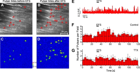
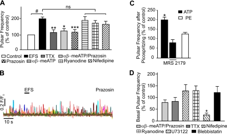
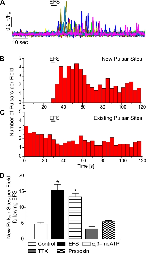
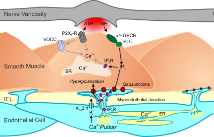
It is generally accepted that the endothelium regulates vascular tone independent of the activity of the sympathetic nervous system. Here, we tested the hypothesis that the activation of sympathetic nerves engages the endothelium to oppose vasoconstriction. Local inositol 1,4,5-trisphosphate (IP(3))-mediated Ca(2+) signals ("pulsars") in or near endothelial projections to vascular smooth muscle (VSM) were measured in an en face mouse mesenteric artery preparation. Electrical field stimulation of sympathetic nerves induced an increase in endothelial cell (EC) Ca(2+) pulsars, recruiting new pulsar sites without affecting activity at existing sites. This increase in Ca(2+) pulsars was blocked by bath application of the α-adrenergic receptor antagonist prazosin or by TTX but was unaffected by directly picospritzing the α-adrenergic receptor agonist phenylephrine onto the vascular endothelium, indicating that nerve-derived norepinephrine acted through α-adrenergic receptors on smooth muscle cells. Moreover, EC Ca(2+) signaling was not blocked by inhibitors of purinergic receptors, ryanodine receptors, or voltage-dependent Ca(2+) channels, suggesting a role for IP(3), rather than Ca(2+), in VSM-to-endothelium communication. Block of intermediate-conductance Ca(2+)-sensitive K(+) channels, which have been shown to colocalize with IP(3) receptors in endothelial projections to VSM, enhanced nerve-evoked constriction. Collectively, our results support the concept of a transcellular negative feedback module whereby sympathetic nerve stimulation elevates EC Ca(2+) signals to oppose vasoconstriction.


Figures

Fig. 1.
Fig. 1.
Stimulation of nerves increases Ca2+ pulsar frequency in the endothelium of third-order mesenteric arteries from connexin40Bac-GCaMP2 mice. A and B: en face arterial preparations. The plus signs indicate pulsar sites in a field over 20 s before (A) and after (B) electrical field stimulation (EFS). C and D: pseudocolor images showing the initiation sites of Ca2+ pulsars (corresponding to the composite images in A and B) 20 s before (C) and after (D) EFS. The color-coded scale represents the fractional fluorescence (F/F0). E: representative fluorescence (F/F0) traces illustrating the effect of EFS on Ca2+ pulsars originating from pulsar sites. F and G: histograms of before, during, and after EFS under control conditions (n = 16 fields, 8 arteries; F) and in the presence of 1 μM TTX (n = 9 fields, 5 arteries; G). Each bar includes SEs and corresponds to pulsars per field during 4 s. The dashed lines indicate the mean frequency before stimulation.
Fig. 2.
Fig. 2.
Inhibition of α-adrenergic receptors prevents EFS-induced increases in Ca2+ pulsar frequency. A: summary of the effects of various agents on EFS-induced Ca2+ pulsar frequency, comparing the 20-s interval starting immediately after the end of the EFS pulse with the 20-s interval before EFS (control). Events that occurred during EFS were excluded from this analysis because some images showed excessive movement during EFS. EFS significantly increased pulsar activity compared with unstimulated controls. This increase in EC Ca2+ pulsars was prevented by pretreatment with 1 μM TTX (n = 8), 500 nM prazosin/10 μM αβ-meATP (n = 6), or 500 nM prazosin alone (n = 18), which reduced pulsar frequency to levels that were not significantly different from unstimulated controls. EFS-induced pulsar activity was not significantly changed by treatment with 10 μM αβ-meATP (n = 10), 10 μM ryanodine (n = 6), or 5 μM nifedipine (n = 5). ns, Not significant. #P < 0.01 vs. unstimulated controls; *P < 0.05, **P < 0.001, and ***P < 0.0001 vs. EFS alone. B: representative fluorescence (F/F0) traces illustrating the lack of effect of EFS on Ca2+ pulsar activity in the presence of prazosin (500 nM). C: summary graph depicting the twofold elevation in pulsar frequency induced by picospritzing 10 μM ATP directly on endothelial cells (ECs) in an en face arterial preparation (n = 3, *P < 0.05). ATP had no effect in the presence of 3 μM MRS-2179 (n = 3), which was routinely included in bath solutions. Picospritzing 100 μM phenylephrine (PE; n = 3) on ECs in an en face preparation did not increase Ca2+ pulsar frequency, indicating that neurally released norepinephrine (NE) does not act directly on the endothelium. D: effects of drug treatments on basal pulsar activity. αβ-meATP (10 μM) + prazosin (500 nM), TTX (1 μM), nifedipine (5 μM), ryanodine (10 μM), and blebbistatin (50 μM) had no significant effects on basal pulsar activity (n = 3–6). Application of 10 μM U-73122 significantly reduced basal pulsar activity (n = 3–4; *P < 0.05).
Fig. 3.
Fig. 3.
Stimulation of sympathetic nerves induces the recruitment of new EC Ca2+ pulsar sites. A: representative traces of the time course of Ca2+ pulsar activity at newly recruited sites after EFS. B and C: histograms of Ca2+ pulsars at newly recruited pulsar sites (n = 11 fields, 5 arteries; B) and preexisting sites (n = 34 fields, 15 arteries; C). D: summary graph showing a significantly increased number of new Ca2+ pulsar sites per field over the course of 20 s after EFS (n = 13, P < 0.0001) compared with the time control in the absence of EFS (n = 3). The number of new Ca2+ pulsar sites after EFS was also significantly increased in the presence of 10 μM αβ-meATP (n = 27, *P < 0.0001) but not in the presence of 1 μM TTX (n = 12) or 500 nM prazosin (n = 21).
Fig. 4.
Fig. 4.
Intermediate-conductance (IK) channels oppose nerve-induced constriction via a mechanism that is dependent on vascular smooth muscle cell α-adrenergic receptor activation and independent of nitric oxide synthase/cyclooxygenase. A–E: representative diameter changes in pressurized (80 mmHg, 166 ± 25-μm maximal diameter and 129 ± 23-μm resting diameter, n = 34 arteries) third-order mesenteric arteries in response to EFS (indicated by arrows). Gray areas indicate area under the curve, which was calculated as an integral of the constriction. A: application of 100 nM charybdotoxin (ChTx) in the presence of 1 μM paxilline (Pax) increased constriction to identical nerve stimulation by twofold (n = 6 arteries). B: application of 300 nM apamin did not cause vasoconstriction (1.4 ± 1.0%, n = 5 arteries) and did not significantly alter nerve-induced vasoconstriction (P > 0.05, n = 5 arteries). This increase in EFS-induced constriction produced by inhibition of IK channels with ChTx was not significantly effected by pretreatment with 100 μM N-nitro-l-arginine methyl ester (l-NAME)-10 μM indomethacin-1 μM Pax (n = 5 arteries; P > 0.05; C) or 10 μM αβ-meATP-1 μM Pax (n = 5 arteries, P > 0.05; D) but was abolished by pretreatment with 500 nM prazosin-1 μM Pax (n = 5 arteries, *P < 0.05; E). Although 10 μM αβ-meATP completely abolished peak constriction in response to EFS, its effects on the area under the curve were minimal. F: summary graph illustrating the changes in luminal diameter upon EFS. For all experiments with ChTx, arteries were first incubated with 1 μM Pax to block smooth muscle large-conductance channels. Control corresponds to pretreatment of vessels with the indicated pathway inhibitors (in the presence of Pax).
Fig. 5.
Fig. 5.
Model of negative feedback regulation by the endothelium in response to sympathetic nerve stimulation. Activation of sympathetic nerves leads to the corelease of ATP and NE, which activate vascular smooth muscle cell purinergic receptors (P2X1Rs) and α1-adrenergic receptors [α1-G protein-coupled receptors (α1-GPCR)], respectively. The subsequent elevation of smooth muscle inositol 1,4,5-trisphosphate (IP3) by the activation of α1-adrenergic receptors is transmitted through myoendothelial gap junctions to IP3 receptors (IP3Rs) in the endothelial projections to increase intracellular Ca2+ (pulsars). This, in turn, activates colocalized EC IK channels, providing a hyperpolarizing influence on the smooth muscle to oppose vasoconstriction. VDCC, voltage-dependent Ca2+ channels; PLC, phospholipase C; SR, sarcoplasmic reticulum; IEL, inner elastic lamina; ER, endoplasmic reticulum.

References

    1. Allbritton NL, Meyer T, Stryer L. Range of messenger action of calcium ion and inositol 1,4,5-trisphosphate. Science 258: 1812–1815, 1992 - PubMed
    1. Burnham MP, Bychkov R, Feletou M, Richards GR, Vanhoutte PM, Weston AH, Edwards G. Characterization of an apamin-sensitive small-conductance Ca2+-activated K+ channel in porcine coronary artery endothelium: relevance to EDHF. Br J Pharmacol 135: 1133–1143, 2002 - PMC - PubMed
    1. Bychkov R, Burnham MP, Richards GR, Edwards G, Weston AH, Feletou M, Vanhoutte PM. Characterization of a charybdotoxin-sensitive intermediate conductance Ca2+-activated K+ channel in porcine coronary endothelium: relevance to EDHF. Br J Pharmacol 137: 1346–1354, 2002 - PMC - PubMed
    1. Dora KA, Doyle MP, Duling BR. Elevation of intracellular calcium in smooth muscle causes endothelial cell generation of NO in arterioles. Proc Natl Acad Sci USA 94: 6529–6534, 1997 - PMC - PubMed
    1. Feletou M, Huang Y, Vanhoutte PM. Endothelium-mediated control of vascular tone: COX-1 and COX-2 products. Br J Pharmacol 164: 894–912, 2011 - PMC - PubMed

Publication types

MeSH terms

Substances

LinkOut - more resources