. 2012 Feb 15;302(4):L370-9.
doi: 10.1152/ajplung.00349.2011. Epub 2011 Dec 2.

Activation of calpains mediates early lung neutrophilic inflammation in ventilator-induced lung injury

Affiliations

Activation of calpains mediates early lung neutrophilic inflammation in ventilator-induced lung injury

Dejie Liu et al. Am J Physiol Lung Cell Mol Physiol. .

Abstract

Lung inflammatory responses in the absence of infection are considered to be one of primary mechanisms of ventilator-induced lung injury. Here, we determined the role of calpain in the pathogenesis of lung inflammation attributable to mechanical ventilation. Male C57BL/6J mice were subjected to high (28 ml/kg) tidal volume ventilation for 2 h in the absence and presence of calpain inhibitor I (10 mg/kg). To address the isoform-specific functions of calpain 1 and calpain 2 during mechanical ventilation, we utilized a liposome-based delivery system to introduce small interfering RNAs targeting each isoform in pulmonary vasculature in vivo. Mechanical ventilation with high tidal volume induced rapid (within minutes) and persistent calpain activation and lung inflammation as evidenced by neutrophil recruitment, production of TNF-α and IL-6, pulmonary vascular hyperpermeability, and lung edema formation. Pharmaceutical calpain inhibition significantly attenuated these inflammatory responses caused by lung hyperinflation. Depletion of calpain 1 or calpain 2 had a protective effect against ventilator-induced lung inflammatory responses. Inhibition of calpain activity by means of siRNA silencing or pharmacological inhibition also reduced endothelial nitric oxide (NO) synthase (NOS-3)-mediated NO production and subsequent ICAM-1 phosphorylation following high tidal volume ventilation. These results suggest that calpain activation mediates early lung inflammation during ventilator-induced lung injury via NOS-3/NO-dependent ICAM-1 phosphorylation and neutrophil recruitment. Inhibition of calpain activation may therefore provide a novel and promising strategy for the prevention and treatment of ventilator-induced lung injury.


Figures
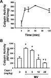
Fig. 1.
Fig. 1.
Effects of mechanical ventilation (MV) and calpain inhibitor (Capn I) on calpain activity in mouse lungs. Calpain activity was measured as described in materials and methods. A: mechanical ventilation increased pulmonary calpain activity. Mice were ventilated with high tidal volume (28 ml/kg) for the indicated time (n = 6 for each time point). B: effects of different concentrations of Capn I on calpain activity. Mice were pretreated with various doses of Capn I (n = 6 per group/dose) 1 h before exposure to high tidal volume MV for 2 h. *P <0.05 vs. control group (normal tidal volume ventilation in the absence of Capn I). †P <0.05 vs. MV control group. RFU, relative fluorescence units.
Fig. 2.
Fig. 2.
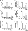
Calpain inhibitor attenuated mechanical ventilation-induced lung injury in mice. Mice were ventilated with high tidal volume (MV) for 2 h in the absence and presence of Capn I (10 mg/kg ip), n = 6–8/group. Normal tidal volume ventilation was used in the control group (CON). A: extravascular lung water (ELW). B: protein concentrations in bronchoalveolar lavage (BAL) fluid. C: polymorphonuclear neutrophil (PMN) counts in BAL fluid. D: lung myeloperoxidase (MPO) activity. E and F: levels of TNF-α (E) and IL-6 (F) in BAL fluid. *P < 0.05 vs. CON groups. †P <0.05 vs. MV groups.
Fig. 3.
Fig. 3.
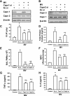
Decreased lung inflammation and injury by MV in calpain-depleted mice. Mice were transfected with scrambled (Sc) and calpain 1 (Capn1) or calpain 2 (Capn2) siRNA. After 48 h, mice (n = 6/group) were ventilated with high tidal volume (28 ml/kg) for 2 h. Sc siRNA-treated (Sc si) mice ventilated with normal tidal volume were used in the control groups. Effective knockdown of calpain 1 and calpain 2 expressions was confirmed by Western blotting of lung homogenates. A and B: calpain 1 (A) and calpain 2 (B) protein expressions were specifically downregulated by calpain 1 siRNA and calpain 2 siRNA, respectively. Results are representative of 3 independent experiments. C: ELW, n = 6. D: protein concentrations in BAL fluid (n = 6). E: PMN counts in BAL fluid. F: lung MPO activity (n = 6). G and H: levels of TNF-α (G) and IL-6 (H) in BAL fluid. *P < 0.05 vs. control groups. †P <0.05 vs. Sc siRNA + MV groups.
Fig. 4.
Fig. 4.
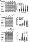
Effects of calpain inhibitor and depletion of calpain on MV-induced ICAM-1 phosphorylation in mouse lungs. Left: representative Western blots of protein expression. All experiments were repeated at least 3 times. Right: density of proteins and phosphoproteins in respective control group was used as a standard (1 arbitrary unit) to compare relative densities in the other groups. A: time course of mechanical ventilation-induced ICAM-1 phosphorylation at Tyr518 in lungs. Mice were ventilated with high tidal volume (28 ml/kg) for the indicated time. B: effects of calpain inhibitor on ICAM-1 phosphorylation attributable to mechanical ventilation. Mice were ventilated with high tidal volume (MV, 28 ml/kg) for 2 h in the absence and presence of Capn I (10 mg/kg ip). Normal tidal volume ventilation was used in the control group. C: effect of depletion of calpain in pulmonary vasculature on ICAM-1 phosphorylation in lungs. Mice were transfected with scrambled, calpain 1, or calpain 2 siRNA. After 48 h, mice were ventilated with high tidal volume (28 ml/kg) for 2 h. Sc siRNA-treated mice ventilated with normal tidal volume were used in the control groups. *P < 0.05 vs. control groups. †P <0.05 vs. MV (B) and Sc siRNA + MV (C) groups.
Fig. 5.
Fig. 5.
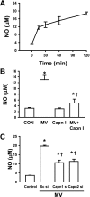
Effects of calpain inhibitor and depletion of calpain on nitric oxide (NO) synthase-3-derived NO production in mouse lungs following mechanical ventilation. NO production was measured as described in materials and methods. A: time course of mechanical ventilation-induced NO production in lungs. Mice were ventilated with high tidal volume (28 ml/kg) for the indicated time (n = 4–6 per each time point). B: effects of calpain inhibitor on NO production attributable to mechanical ventilation. Mice (n = 6) were ventilated with high tidal volume (MV, 28 ml/kg) for 2 h in the absence and presence of Capn I (10 mg/kg ip). Normal tidal volume ventilation was used in the control group. C: effects of depletion of calpain in pulmonary vasculature on NO production in lungs. Mice were transfected with scrambled and calpain 1 or calpain 2 siRNA. After 48 h, mice (n = 6/group) were ventilated with high tidal volume (28 ml/kg) for 2 h. Sc siRNA-treated mice ventilated with normal tidal volume were used in the control groups. *P < 0.05 vs. control groups. †P <0.05 vs. MV (B) and Sc siRNA + MV (C) groups.
Fig. 6.
Fig. 6.
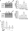
NO induced ICAM-1 phosphorylation and neutrophil recruitment into the mouse lung. A: effects of NO synthase (NOS) inhibitor on ICAM-1 phosphorylation attributable to mechanical ventilation. Mice were ventilated with high tidal volume (MV, 28 ml/kg) for 2 h in the absence and presence of NOS inhibitor nitro-l-arginine methyl ester (l-NAME) (15 mg/kg ip). Normal tidal volume ventilation was used in the control group. Left: representative Western blots of protein expression (n = 3). Right: density of proteins and phosphoproteins in respective control group was used as a standard (1 arbitrary unit) to compare relative densities in the other groups. B: effects of tyrosine inhibitor genistein (GENI) on ICAM-1 phosphorylation in lungs. Mice were ventilated with high tidal volume (MV, 28 ml/kg) for 2 h in the absence and presence of genistein (50 mg/kg ip). Normal tidal volume ventilation was used in the control group. Left: representative Western blots of protein expression. Right: density of proteins and phosphoproteins in respective control group was used as a standard (1 arbitrary unit) to compare relative densities in the other groups (n = 3). C: effects of NOS and tyrosine inhibitor on PMN infiltration into the lung (n = 6/group). *P < 0.05 vs. control groups. †P <0.05 vs. MV alone group.
Fig. 7.
Fig. 7.
Effects of calpain inhibitor and depletion of calpain on mechanical ventilation-induced NOS-2 expression, and phosphorylation of NOS-3 and Akt in mouse lungs. Protein expressions of NOS-3, NOS-2, and Akt as well as phosphorylation of NOS-3 and Akt were determined by Western blot analysis. All experiments were repeated at least 3 times. Left: representative Western blots of protein expression. Right: density of proteins and phosphoproteins in respective control group was used as a standard (1 arbitrary unit) to compare relative densities in the other groups. A: time course of mechanical ventilation-induced NOS-3, NOS-2, Akt protein expressions, and phosphorylation of NOS-3 and Akt in lungs. Mice were ventilated with high tidal volume (28 ml/kg) for the indicated time. B: effects of calpain inhibitor on NOS-3, NOS-2 protein expressions, and phosphorylation of NOS-3 and Akt attributable to MV. Mice were ventilated with high tidal volume (MV, 28 ml/kg) for 2 h in the absence and presence of Capn I (10 mg/kg ip). Normal tidal volume ventilation was used in the control group. C: effects of depletion of calpain in pulmonary vasculature on NOS-3, NOS-2, Akt protein expressions, and phosphorylation of NOS-3 and Akt in lungs. Mice were transfected with scrambled, calpain 1, or calpain 2 siRNA. After 48 h, mice were ventilated with high tidal volume (MV, 28 ml/kg) for 2 h. Sc siRNA-treated mice ventilated with normal tidal volume were used in the control groups. *P < 0.05 vs. control groups. †P <0.05 vs. 60 min (A), MV (B), and Sc siRNA + MV (C) groups.
Fig. 8.
Fig. 8.
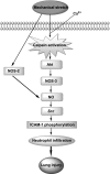
Model of calpains in mediating lung neutrophilic inflammation induced by mechanical stretch.

Similar articles

Cited by

References

    1. Abraham E, Carmody A, Shenkar R, Arcaroli J. Neutrophils as early immunologic effectors in hemorrhage- or endotoxemia-induced acute lung injury. Am J Physiol Lung Cell Mol Physiol 279: L1137–L1145, 2000 - PubMed
    1. Acute Respiratory Distress Syndrome Network Ventilation with lower tidal volumes as compared with traditional tidal volumes for acute lung injury and the acute respiratory distress syndrome. N Engl J Med 342: 1301–1308, 2000 - PubMed
    1. Arthur JS, Elce JS, Hegadorn C, Williams K, Greer PA. Disruption of the murine calpain small subunit gene, Capn4: calpain is essential for embryonic development but not for cell growth and division. Mol Cell Biol 20: 4474–4481, 2000 - PMC - PubMed
    1. Averna M, Stifanese R, De Tullio R, Salamino F, Bertuccio M, Pontremoli S, Melloni E. Proteolytic degradation of nitric oxide synthase isoforms by calpain is modulated by the expression levels of HSP90. FEBS J 274: 6116–6127, 2007 - PubMed
    1. Azam M, Andrabi SS, Sahr KE, Kamath L, Kuliopulos A, Chishti AH. Disruption of the mouse μ-calpain gene reveals an essential role in platelet function. Mol Cell Biol 21: 2213–2220, 2001 - PMC - PubMed

Publication types

MeSH terms

LinkOut - more resources