. 2012 Mar 1;302(5):L463-73.
doi: 10.1152/ajplung.00099.2011. Epub 2011 Dec 2.

Regulation of alveolar epithelial cell apoptosis and pulmonary fibrosis by coordinate expression of components of the fibrinolytic system

Affiliations

Regulation of alveolar epithelial cell apoptosis and pulmonary fibrosis by coordinate expression of components of the fibrinolytic system

Yashodhar P Bhandary et al. Am J Physiol Lung Cell Mol Physiol. .

Abstract

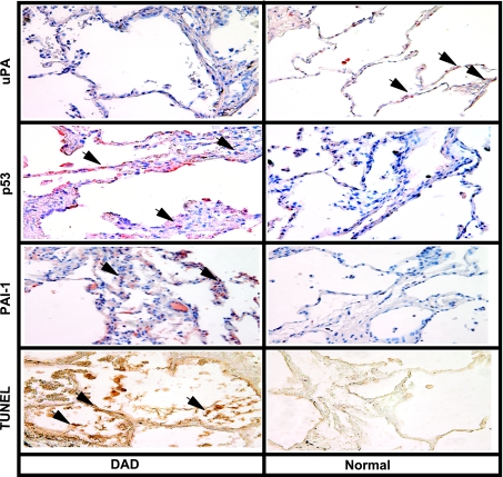
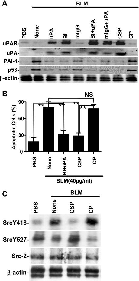
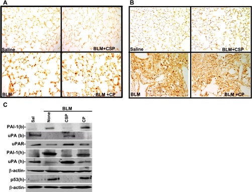
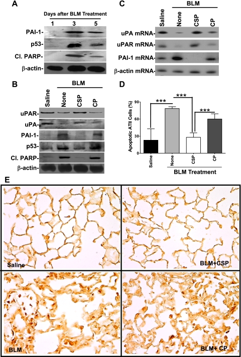
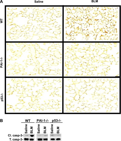
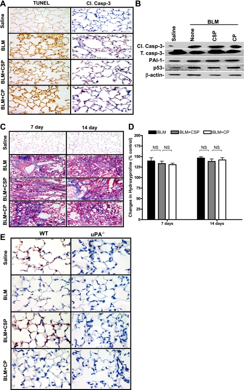
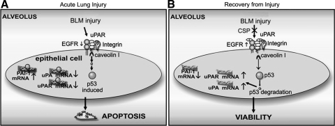
Alveolar type II (ATII) cell apoptosis and depressed fibrinolysis that promotes alveolar fibrin deposition are associated with acute lung injury (ALI) and the development of pulmonary fibrosis (PF). We therefore sought to determine whether p53-mediated inhibition of urokinase-type plasminogen activator (uPA) and induction of plasminogen activator inhibitor-1 (PAI-1) contribute to ATII cell apoptosis that precedes the development of PF. We also sought to determine whether caveolin-1 scaffolding domain peptide (CSP) reverses these changes to protect against ALI and PF. Tissues as well as isolated ATII cells from the lungs of wild-type (WT) mice with BLM injury show increased apoptosis, p53, and PAI-1, and reciprocal suppression of uPA and uPA receptor (uPAR) protein expression. Treatment of WT mice with CSP reverses these effects and protects ATII cells against bleomycin (BLM)-induced apoptosis whereas CSP fails to attenuate ATII cell apoptosis or decrease p53 or PAI-1 in uPA-deficient mice. These mice demonstrate more severe PF. Thus p53 is increased and inhibits expression of uPA and uPAR while increasing PAI-1, changes that promote ATII cell apoptosis in mice with BLM-induced ALI. We show that CSP, an intervention targeting this pathway, protects the lung epithelium from apoptosis and prevents PF in BLM-induced lung injury via uPA-mediated inhibition of p53 and PAI-1.


Figures

Fig. 1.
Fig. 1.
p53, urokinase-type plasminogen activator (uPA), and plasminogen activator inhibitor-1 (PAI-1) expression in the human diffuse alveolar damage (DAD) tissues. Sections from DAD and normal lungs were subjected to immunohistochemistry (IHC) analyses using anti-p53, anti-uPA, or anti-PAI-1 monoclonal antibodies (1:50 dilution) or terminal deoxynucleotidyl transferase dUTP-mediated nick-end labeling (TUNEL) staining to assess the changes in lung epithelial (LE) cell uPA, p53, and PAI-1 expression as well as LE cell apoptosis. Representative fields from 1 of 3 sections per subject are shown at ×200 magnification. Arrows indicate foci of strong staining in each of the representative fields.
Fig. 2.
Fig. 2.
Caveolin-1 scaffolding domain peptide (CSP) reverses bleomycin (BLM)-induced changes in alveolar type II (ATII) cell uPA, uPA receptor (uPAR), PAI-1, and p53 in vitro. A: mouse ATII cells were treated for 28 h at 37°C with PBS or 40 μg/ml of BLM in the presence or absence of either full-length uPA or the aminoterminal fragment (ATF) of uPA (each 20 nM), 1 μg/ml each of anti-β1-integrin monoclonal antibody (BI) or mouse IgG (mIgG), BI+uPA, mIgG+uPA, CSP (10 nM), and control peptide (CP; 10 nM) at 37°C. The membrane proteins were immunoblotted with anti-uPAR antibody. In the case of uPA and PAI-1, the full-length uPA stimulus was substituted with ATF. The conditioned media were analyzed for uPA and PAI-1 while cell lysates were analyzed for p53 and β-actin by Western blotting. Experiments were repeated at least twice and representative results are illustrated. B: inhibition of BLM-induced apoptosis of ATII cells by anti-β1-integrin antibody and uPA or CSP. Mouse ATII cells treated as described above (A) were detached from the culture plates by using trypsin-EDTA solution. These cells were stained with anti-annexin-V antibody-propidium iodide (PI) solution and analyzed for apoptosis by flow cytometry. The percentage of apoptotic cells is presented as bars representing mean ± SD of 4 replications. Differences between treatments are statistically significant (**P < 0.001) or not statistically significant (NS) as confirmed by Student's t-test or 1-way ANOVA. C: CSP inhibits BLM-induced Src activation. Lysates of ATII cells treated with PBS or BLM with or without CSP or CP for 24 h were tested for tyrosine (Y418) phosphorylation and inhibitory tyrosine (Y527) phosphorylation by Western blotting using phosphospecific antibodies. The same membrane was then stripped and tested for total Src (Src-2) kinase and β-actin expression. Doublets found in bottom panels of A and C are due to the recognition of both β- and γ-actins by anti-β-actin antibody.
Fig. 3.
Fig. 3.
CSP inhibits BLM-induced ATII cell apoptosis and alters uPA, uPAR, p53, and PAI-1 in mouse lungs. Mice exposed to BLM were then treated with CSP (18.75 mg/kg body wt) or CP by intranasal instillation on days 2, 4, and 6 after BLM treatment. Lung sections of mice euthanized 7 (A) and 21 (B) days after BLM injury were analyzed for apoptosis by TUNEL staining. TUNEL-stained sections (magnification ×200) are representative of 9 fields/mouse from each of 3 mice. C: bronchoalveolar lavage (BAL) fluids (b) and lung homogenates (h) of mice at 1 wk after BLM injury as described in A were analyzed for uPA, p53, PAI-1, and β-actin proteins by Western blotting. Membrane proteins were immunoblotted for uPAR expression by using mouse-specific antibodies. Results are representative of at least 3 independent repetitions.
Fig. 4.
Fig. 4.
CSP prevents BLM-induced collagen deposition in mouse lungs. A: lung sections of mice euthanized on day 21 after exposure to BLM as described in Fig. 3 were subjected to trichrome staining. Stain indicates the collagen deposition. Figure (magnification ×200) is representative of 9 fields/mouse; n = 3 mice. B: inhibition of lung fibrosis by treatment with CSP in mice. Mice exposed to BLM were implanted with a 0.1-ml microosmotic pump filled with 125 mg/kg body wt of CSP (BLM+CSP) or CP (BLM+CP) on 2 or 7 or 14 days after initiation of injury. The lung homogenates from mice euthanized 21 days after BLM injury were analyzed for hydroxyproline content. Bars represent mean ± SD of at least 3 repetitions (n = 3 mice/group). Differences between treatments are statistically significant (**P < 0.05) or (***P < 0.01) as confirmed by Student's t-test or 1-way ANOVA. C: CSP restores lung functions after BLM injury. BLM-treated mice were intraperitoneally (IP) injected with or without 125 mg/kg body wt of CSP or CP on days 2, 4, and 6 after initial injury. These mice were tested for changes in lung compliance (i) and resistance (ii) to assess respiratory function 21 days after BLM injury as described in materials and methods. Bars represent means ± SD of at least 2 repetitions (n = 3 mice/group). Significant differences (***P < 0.005 and *P < 0.05) in lung compliance or resistance between different experimental groups were confirmed by Student's t-test or 1-way ANOVA.
Fig. 5.
Fig. 5.
CSP reverses BLM-induced changes in ATII cell uPA, uPAR, p53, and PAI-1 expression and apoptosis in vivo. A: ATII cells were isolated from mice on day 1, 3, or 5 after BLM injury and were immunoblotted for PAI-1 and p53 expression and cleaved poly(ADP-ribose) polymerase (Cl.PARP). The same membrane was later stripped and analyzed for β-actin. B: mice exposed to saline or BLM for 24 h were given IP injection of CSP or CP. ATII cells extracted from the lungs of these mice 3 days after BLM injury were analyzed for uPA, uPAR, p53, PAI-1, Cl.PARP, and β-actin by Western blotting. C: total RNA extracted ATII cells from mice treated as described in B were tested for changes or uPA, uPAR, PAI-1, and β-actin mRNA by RT-PCR. D: ATII cells isolated from mice as described in B were treated with anti-annexin-V antibody-PI solution and subjected to flow cytometry. The percentage of apoptotic cells is presented as a bar graph representing mean ± SD of 4 replications. Significant differences (***P < 0.001) between the groups were confirmed by Student's t-test or 1-way ANOVA. E: mice exposed to BLM were treated with or without CSP or CP 24 h after BLM injury. Lung sections from these mice euthanized 3 days after initial BLM injury and control mice were analyzed for apoptosis by TUNEL staining.
Fig. 6.
Fig. 6.
p53- and PAI-1-deficient mice resist ATII cell apoptosis. A: lung sections of wild-type (WT), p53-, and PAI-1-deficient mice euthanized 3 days after the exposure to saline or BLM were subjected to TUNEL staining. The stained sections (magnification ×200) are representative of 9 fields/mouse (n = 3 mice). B: ATII cells isolated from WT and p53- and PAI-1-deficient mice 3 days after the exposure to saline or BLM were analyzed for active (Cl.) and total (T) caspase-3 (casp-3) by Western blotting to assess apoptosis.
Fig. 7.
Fig. 7.
uPA is involved in CSP-mediated protection against BLM-induced apoptosis of ATII cells and prevention of PF. A: lung sections of uPA-deficient mice exposed to BLM with or without CSP or CP euthanized 3 days after BLM injury were tested for apoptosis by TUNEL staining and IHC using an antibody that detects Cl.caspase-3 (1:50 dilution). Mice exposed to saline were used as controls. Representative sections from 3 mice are shown at ×400 magnification. B: ATII cells isolated 72 h after BLM injury from uPA-deficient mice treated with saline, BLM, BLM+CSP or BLM+CP as described in A were analyzed for Cl./T. caspase-3, PAI-1, p53, and β-actin proteins by Western blotting. C: 7 or 14 days after BLM injury, uPA-deficient mice were IP injected with or without CSP or CP. Saline-treated mice were used as controls. Lung sections from these mice euthanized 21 days after BLM injury were subjected to trichrome staining. Sections representative of 9 fields/mouse (n = 3 mice) are shown at ×200 magnification. D: lung homogenates from mice treated as described in C were analyzed for the changes in total hydroxyproline contents. Bars represent means ± SD of at least 3 repetitions (n = 3 mice/group). Significant differences between treatment and control groups were confirmed by Student's t-test or 1-way ANOVA. E: representative lung sections of WT and uPA-deficient mice exposed to BLM with or without CSP or CP euthanized 3 days after BLM injury (n = 3 mice/group) were stained for the proliferation marker Ki-67 by using an anti-Ki-67 antibody (1:50 dilution). Mice exposed to saline were used as controls. Representative sections from 3 mice are shown at ×400 magnification.
Fig. 8.
Fig. 8.
Regulation of LE cell viability through cross talk between p53 and components of fibrinolytic system. A: p53 is induced during acute lung injury then binds transcripts for uPA, uPAR, and PAI-1 and inhibits uPA and uPAR mRNA through destabilization of the transcripts (46, 54). PAI-1 is induced through p53-mediated stabilization of PAI-1 mRNA (55); the end result is reciprocal suppression of uPA and uPAR and increased PAI-1 expression by LE cells. These conditions result in enhanced LE cell apoptosis. B: expression of p53 can be inhibited by activation of the β1-integrin-uPAR-EGFR complex through CSP, resulting in competitive inhibition of caveolin-1. The paucity of p53 in LE cells prevents p53 binding to uPA, uPAR, and PAI-1 mRNA, so uPA and uPAR transcripts remain stable whereas PAI-1 mRNA is destabilized. uPA and uPAR levels in LE cells are preserved and serve as survival signals whereas PAI-1 expression is inhibited.

References

    1. Alfano D, Franco P, Vocca I, Gambi N, Pisa V, Mancini A, Caputi M, Carriero MV, Iaccarino I, Stoppelli MP. The urokinase plasminogen activator and its receptor: role in cell growth and apoptosis. Thromb Haemost 93: 205– 211, 2005 - PubMed
    1. Barazzone C, Belin D, Piguet PF, Vassalli JD, Sappino AP. Plasminogen activator inhibitor-1 in acute hyperoxic mouse lung injury. J Clin Invest 98: 2666– 2673, 1996 - PMC - PubMed
    1. Barker SC, Kassel DB, Weigl D, Huang X, Luther MA, Knight WB. Characterization of pp60c-src tyrosine kinase activities using a continuous assay: autoactivation of the enzyme is an intermolecular autophosphorylation process. Biochemistry 34: 14843– 14851, 1995 - PubMed
    1. Bertozzi P, Astedt B, Zenzius L, Lynch K, LeMaire F, Zapol W, Chapman HA., Jr Depressed bronchoalveolar urokinase activity in patients with adult respiratory distress syndrome. N Engl J Med 322: 890– 897, 1990 - PubMed
    1. Bhandary YP, Shetty SK, Shetty R, Idell S, Starcher B, Shetty S. Regulation of lung epithelial apoptosis by coordinate expression of components of the fibrinolytic system (Abstract). Am J Respir Crit Care Med 181: A4185, 2010

Publication types

MeSH terms