

Structural insights into the Cdt1-mediated MCM2-7 chromatin loading
- PMID: 22140117
- PMCID: PMC3326298
- DOI: 10.1093/nar/gkr1118
Structural insights into the Cdt1-mediated MCM2-7 chromatin loading
Abstract
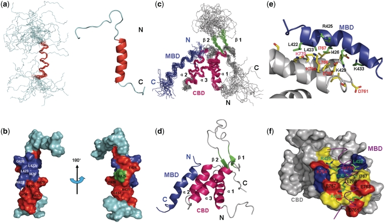
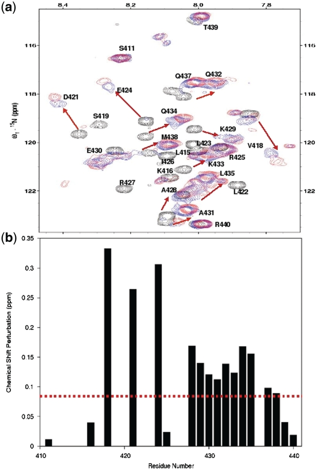
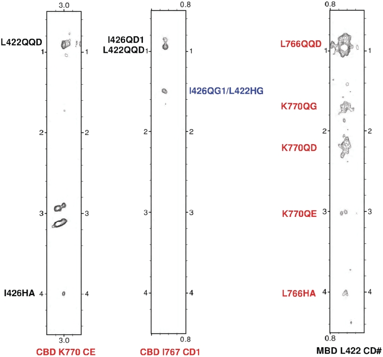
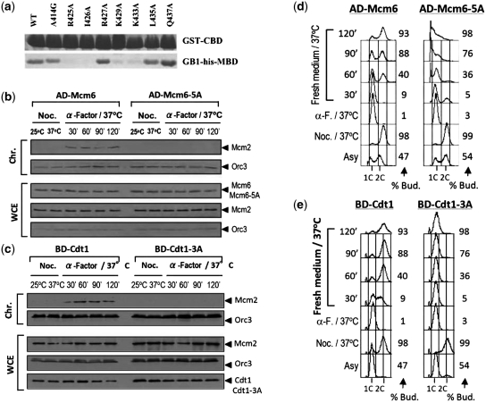
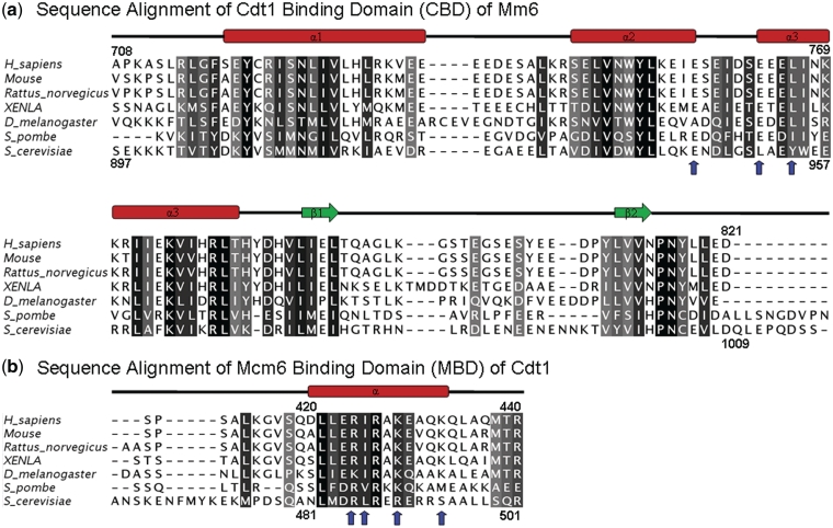
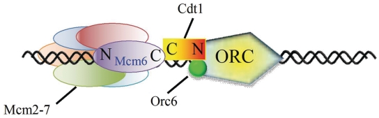
Initiation of DNA replication in eukaryotes is exquisitely regulated to ensure that DNA replication occurs exactly once in each cell division. A conserved and essential step for the initiation of eukaryotic DNA replication is the loading of the mini-chromosome maintenance 2-7 (MCM2-7) helicase onto chromatin at replication origins by Cdt1. To elucidate the molecular mechanism of this event, we determined the structure of the human Cdt1-Mcm6 binding domains, the Cdt1(410-440)/MCM6(708-821) complex by NMR. Our structural and site-directed mutagenesis studies showed that charge complementarity is a key determinant for the specific interaction between Cdt1 and Mcm2-7. When this interaction was interrupted by alanine substitutions of the conserved interacting residues, the corresponding yeast Cdt1 and Mcm6 mutants were defective in DNA replication and the chromatin loading of Mcm2, resulting in cell death. Having shown that Cdt1 and Mcm6 interact through their C-termini, and knowing that Cdt1 is tethered to Orc6 during the loading of MCM2-7, our results suggest that the MCM2-7 hexamer is loaded with its C terminal end facing the ORC complex. These results provide a structural basis for the Cdt1-mediated MCM2-7 chromatin loading.
Figures
Similar articles
-
Structural basis of Mcm2-7 replicative helicase loading by ORC-Cdc6 and Cdt1.Nat Struct Mol Biol. 2017 Mar;24(3):316-324. doi: 10.1038/nsmb.3372. Epub 2017 Feb 13. Nat Struct Mol Biol. 2017. PMID: 28191893 Free PMC article.
-
Structural mechanism of helicase loading onto replication origin DNA by ORC-Cdc6.Proc Natl Acad Sci U S A. 2020 Jul 28;117(30):17747-17756. doi: 10.1073/pnas.2006231117. Epub 2020 Jul 15. Proc Natl Acad Sci U S A. 2020. PMID: 32669428 Free PMC article.
-
1H, 15N and 13C chemical shift assignments of the Cdt1 binding domain of human Mcm6.Biomol NMR Assign. 2010 Oct;4(2):231-3. doi: 10.1007/s12104-010-9246-5. Epub 2010 Jul 11. Biomol NMR Assign. 2010. PMID: 20623209
-
Cdt1 and geminin in DNA replication initiation.Subcell Biochem. 2012;62:71-87. doi: 10.1007/978-94-007-4572-8_5. Subcell Biochem. 2012. PMID: 22918581 Review.
-
Regulation of DNA Replication Licensing and Re-Replication by Cdt1.Int J Mol Sci. 2021 May 14;22(10):5195. doi: 10.3390/ijms22105195. Int J Mol Sci. 2021. PMID: 34068957 Free PMC article. Review.
Cited by
-
Stabilisation of half MCM ring by Cdt1 during DNA insertion.Nat Commun. 2021 Mar 19;12(1):1746. doi: 10.1038/s41467-021-21932-8. Nat Commun. 2021. PMID: 33741931 Free PMC article.
-
Transcriptome Analysis of the Anti-Proliferative Effects of Ginsenoside Rh3 on HCT116 Colorectal Cancer Cells.Molecules. 2022 Aug 6;27(15):5002. doi: 10.3390/molecules27155002. Molecules. 2022. PMID: 35956952 Free PMC article.
-
Characterization of conserved arginine residues on Cdt1 that affect licensing activity and interaction with Geminin or Mcm complex.Cell Cycle. 2016 May 2;15(9):1213-26. doi: 10.1080/15384101.2015.1106652. Cell Cycle. 2016. PMID: 26940553 Free PMC article.
-
Mechanisms and regulation of DNA replication initiation in eukaryotes.Crit Rev Biochem Mol Biol. 2017 Apr;52(2):107-144. doi: 10.1080/10409238.2016.1274717. Epub 2017 Jan 17. Crit Rev Biochem Mol Biol. 2017. PMID: 28094588 Free PMC article. Review.
-
Structural basis of Mcm2-7 replicative helicase loading by ORC-Cdc6 and Cdt1.Nat Struct Mol Biol. 2017 Mar;24(3):316-324. doi: 10.1038/nsmb.3372. Epub 2017 Feb 13. Nat Struct Mol Biol. 2017. PMID: 28191893 Free PMC article.
References
-
- Remus D, Diffley JF. Eukaryotic DNA replication control: lock and load, then fire. Curr. Opin. Cell. Biol. 2009;21:771–777. - PubMed
-
- Bell SP, Dutta A. DNA replication in eukaryotic cells. Annu. Rev. Biochem. 2002;71:333–374. - PubMed
-
- Diffley JFX. Regulation of early events in chromosome replication. Curr. Biol. 2004;14:778–786. - PubMed
-
- Maiorano D, Moreau J, Mechali M. XCDT1 is required for the assembly of pre-replicative complexes in X. laevis. Nature. 2000;404:622–625. - PubMed
-
- Liang C, Weinreich M, Stillman B. ORC and Cdc6p interact and determine the frequency of initiation of DNA replication in the genome. Cell. 1995;81:667–676. - PubMed
Publication types
MeSH terms
Substances
LinkOut - more resources
Full Text Sources
Molecular Biology Databases
Research Materials
Miscellaneous
