. 2012 Mar;60(3):188-204.
doi: 10.1369/0022155411433331. Epub 2011 Dec 1.

YKL-40 is differentially expressed in human embryonic stem cells and in cell progeny of the three germ layers

Affiliations

YKL-40 is differentially expressed in human embryonic stem cells and in cell progeny of the three germ layers

Christian B Brøchner et al. J Histochem Cytochem. 2012 Mar.

Abstract

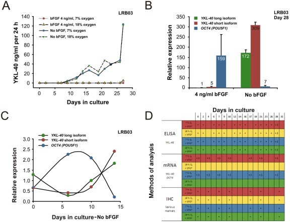
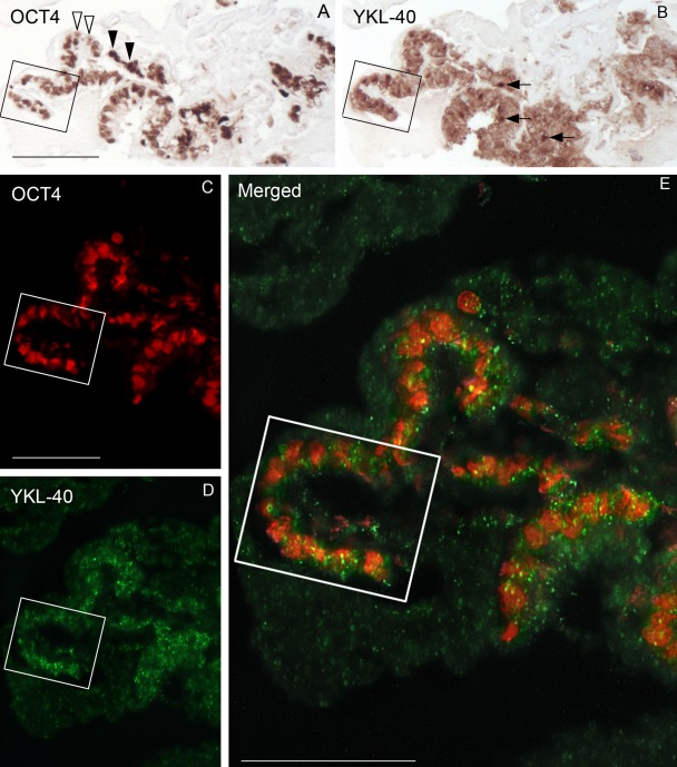
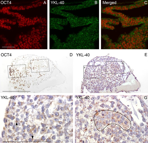
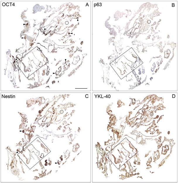
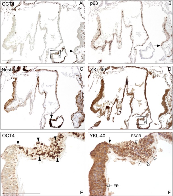
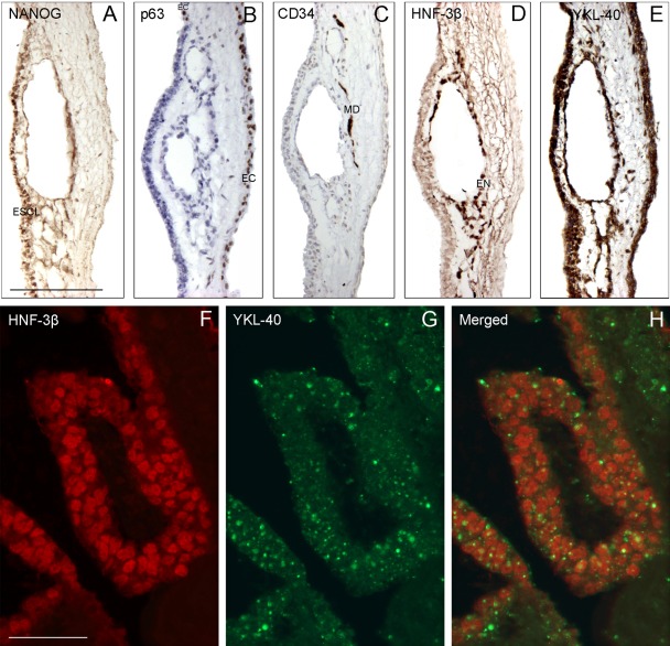
The secreted glycoprotein YKL-40 participates in cell differentiation, inflammation, and cancer progression. High YKL-40 expression is reported during early human development, but its functions are unknown. Six human embryonic stem cell (hESC) lines were cultured in an atmosphere of low or high oxygen tension, in culture medium with or without basic fibroblast growth factor, and on feeder layers comprising mouse embryonic fibroblasts or human foreskin fibroblasts to evaluate whether hESCs and their progeny produced YKL-40 and to characterize YKL-40 expression during differentiation. Secreted YKL-40 protein and YKL-40 mRNA expression were measured by enzyme-linked immunosorbent assay (ELISA) and quantitative RT-PCR. Serial-sectioned colonies were stained for YKL-40 protein and for pluripotent hESC (OCT4, NANOG) and germ layer (HNF-3β, PDX1, CD34, p63, nestin, PAX6) markers. Double-labeling showed YKL-40 expression in OCT4-positive hESCs, PAX6-positive neuroectodermal cells, and HNF-3β-positive endodermal cells. The differentiating progeny showed strong YKL-40 expression. Abrupt transition between YKL-40 and OCT4-positive hESCs and YKL-40-positive ecto- and neuroectodermal lineages was observed within the same epithelial-like layer. YKL-40-positive cells within deeper layers lacked contact with OCT4-positive cells. YKL-40 may be important in initial cell differentiation from hESCs toward ectoderm and neuroectoderm, with retained epithelial morphology, whereas later differentiation into endoderm and mesoderm involves a transition into the deeper layers of the colony.


Conflict of interest statement

The authors declared no potential conflicts of interest with respect to the authorship and/or publication of this article.

Figures

Figure 1.
Figure 1.
Cell line LRB03 grown for up to 32 days. ELISA measurements of YKL-40 protein secretion into the culture medium (A) and mRNA analysis of YKL-40 and OCT4 expression in culture by quantitative RT-PCR (QPCR) (B, C). (A) Concentration of YKL-40 secreted into the culture medium during 28 days of culture, determined by ELISA. Oxygen tension alterations show no difference in amount of YKL-40 measured, whereas absence of basic fibroblast growth factor (bFGF) leads to earlier secretion of YKL-40 into the medium already after 10 days in culture. (B) Relative expression of mRNA for OCT4 and YKL-40 short and long isoforms in relation to culture medium composition with absence (“differentiation medium”) or presence (“proliferation medium”) of 4 ng/ml bFGF, after 28 days of culture. Upregulation of YKL-40 mRNA expression in the human embryonic stem cells (hESCs) cultured without bFGF was 172-fold and 62-fold for the long and short isoforms, respectively, whereas OCT4 was only high when grown with bFGF added. (C) Measurements of OCT4 and both isoforms of YKL-40 by QPCR from culture days 0 to 15 grown without bFGF. YKL-40 isoforms follow each other, leaving no significant change in expression throughout culture age. At culture day 0, OCT4 and both isoforms of YKL-40 mRNA are found in low levels. Shortly thereafter, OCT4 expression rises with a maximum toward day 7 as YKL-40 diminishes. From here on, OCT4 and YKL-40 expression is inversed, which correlates well with the findings of less undifferentiated cells and more differentiated germ layer marker-positive cells. (D) Overview of the different culture conditions employed. Conducted experiments with immunohistochemistry analysis in direct relation to either ELISA or QPCR measurements are shown. •Analysis performed. §Colony terminated. Same color code in (A) and (D) facilitates comparison.
Figure 2.
Figure 2.
Staining patterns for OCT4 (A), YKL-40 (B), and OCT4/YKL-40 (C–E) in three consecutive sections of a human embryonic stem cell colony (LRB03) from culture day 0 visualized by immunohistochemistry and confocal fluorescence microscopy. The “U-turn” in the boxed area is easy to identify in the three sections. (A) All cell nuclei exhibit OCT4 positivity, although a varying intensity of the staining is evident. Open arrowheads point to weakly stained nuclei, and solid arrowheads show strongly labeled nuclei. (B) The adjacent section stained for YKL-40 shows a marked reactivity in the cytoplasm with occasionally denser areas (arrows). (C–E) Confocal microscopy of a neighboring section stained simultaneously with antibodies against OCT4 and YKL-40. Cells with nuclear reactivity (red) for OCT4 (C) also exhibit cytoplasmic reactivity (green) for YKL-40 (D), verified at higher magnification by the merged image (E). Yellow dots correspond to YKL-40 located directly below or above the OCT4-positive red nucleus. Scale bars: 100 µm.
Figure 3.
Figure 3.
Early and later differentiating colonies. (A–C) Confocal microscopy of a 7-day-old colony from LRB03 grown without basic fibroblast growth factor shows early differentiating human embryonic stem cells (hESCs) stained for OCT4 (A) and YKL-40 (B). The merged picture (C) depicts strongly OCT4-reacting stem cell nuclei surrounded by a fine granular cytoplasmic YKL-40 staining. A later differentiating colony (shown in D–G) in bright-field microscopy is from a 17-day-old culture from LRB010 also stained for OCT4 (D) and YKL-40 (E). In (D), a small group of strongly OCT4-positive cells is present toward one edge of the colony with few weakly OCT4-positive cells scattered toward the middle. A neighboring section stained for YKL-40 (E) with boxed areas shown at higher magnification in (F) and (G) depicts YKL-40 in the cytoplasm of loosely packed cells, often in close proximity to the nucleus (arrows in F). Morphology of the individual cells is corresponding well to hESC morphology with large nuclei with one to three dense nucleoli in a scanty cytoplasm. However, the cell pattern is less epithelial and more mesenchymal. In the rather OCT4-negative field in (G), the epithelial-like cell group marked by a dotted line shows a dense granular cytoplasmic YKL-40 reactivity and nuclei without dense nucleoli. M shows a mitotic figure. A–C: same magnification, scale bar: 50 µm. D, E: same magnification, scale bar: 250 µm. F, G: same magnification, scale bar: 100 µm.
Figure 4.
Figure 4.
Survey of entire human embryonic stem cell (hESC) culture from LRB03 embedded at culture day 32 with neighboring sections stained for OCT4 (A), p63 (B), nestin (C), and YKL-40 (D). Boxed areas are shown at higher magnification in Figure 5. (A) Small OCT4-positive areas are distributed widely across the entire culture (arrowheads) in contrast to the vast OCT4-negative areas. (B) Ectodermal marker p63 is positive in well-defined regions of the culture, with a distinct borderline to OCT4 areas (arrows), but this is better appreciated at higher magnification (Fig. 5B). (C) A moderate nestin-positive staining is seen in areas overlapping the OCT4 stained areas (arrowheads). The stronger nestin-reacting subregions of the culture are not OCT4 positive (see also Fig. 5C). (D) YKL-40 is present in all regions and subregions throughout the culture, both in the pluripotent OCT4-positive cells and in the various progenitor cells. YKL-40 reactivity is also seen in structures that show no OCT4, p63, or nestin reactivity (see encircled area). A–D: same magnification, scale bar: 1 mm.
Figure 5.
Figure 5.
(A–D) Higher magnification of boxed areas in Figure 4. Transition points from OCT4-positive cells to p63-positive ectodermal and nestin-positive neuroectodermal cells in the same layer are indicated by single arrows (A, B, C). YKL-40 is positive in areas with pluripotent undifferentiated cells and the more differentiated ectodermal lineages (D). Cells with nuclear OCT4 (dense arrowheads) and cytoplasmic and membranous YKL-40 staining at higher magnification of boxed areas from A and D (E, F). YKL-40 is seen along the membranes (open arrowheads) of the OCT4-positive cells, with pluripotent stem cell morphology in contrast to the strong cytoplasmic reactivity of the adjacent epithelial region (ER). The borderline between the embryonic stem cell region (ESCR) and the epithelial region is indicated by the dense arrow. A–D: same magnification, scale bar: 500 µm. E, F: same magnification, scale bar: 100 µm.
Figure 6.
Figure 6.
(A–E) Markers for human embryonic stem cells (NANOG), ectoderm (p63), mesoderm (CD34), endoderm (HNF-3b), and YKL-40 applied to neighboring sections from an LRB01 colony cultured for 42 days. The p63-positive cells line the outermost area of the culture, whereas the CD34-positive cells are in the middle of the culture. The HNF-3b-positive cells are encircling a tube-like structure in the innermost part of the culture. YKL-40-positive cells are in all three germ layer–like areas as well as in the NANOG-positive cells lining the edge. (F–H) Confocal microscopy of a section from an earlier stage (28 days) of the same LRB01 colony stained for HNF-3b (F) and YKL-40 (G). The merged picture (H) shows the red HNF-3b endodermal cell nuclei surrounded by coarse green granules in the YKL-40-stained cytoplasm. (A–E) Same magnification, scale bar: 200 µm. (F–H) Same magnification, scale bar: 50 µm. ESCL, embryonic stem cell layer; EC, ectodermal layer; MD, mesodermal layer; EN, endodermal layer.
Figure 7.
Figure 7.
(A–E) A series of sections adjacent to those shown in Figure 6A–E (i.e., also 42 days old), stained for markers for undifferentiated stem cells (OCT4); neuroectodermal cells (PAX6); endodermal germ layer (HNF-3β), including the pancreas-duodenum part (PDX1); and YKL-40. A well-defined area consisting of pluripotent OCT4-positive cells is confined to the outermost lining—an embryonic stem cell layer (ESCL), which continues into an ectodermal layer (EC), which extends to a PAX6-positive neuroectoderm (NEC). Cells positive for endodermal germ layer markers (EN) are localized in a deeper layer. The transcription factor PDX1 is seen in the endoderm, differentiating into the pancreas-duodenum anlage (PDEN). YKL-40-positive staining is found in the ESCL, the germ layers, and the PDX1-positive cell aggregate (arrows in E). (F–H) Confocal microscopy of a section from the same series as that shown in A–E, stained for PAX6 (F) and YKL-40 (G). The boxed areas in B and E correspond to F and G (i.e., two entirely different staining protocols lead to very similar results). The merged picture (H) shows the red PAX6-positive neuroectodermal cell nuclei surrounded by a fine granular green YKL-40-positive cytoplasm. Note the stronger YKL-40 staining of the ectoderm. A–E: same magnification, scale bar: 200 µm. F–H: same magnification, scale bar: 20 µm.

Similar articles

Cited by

References

    1. Adewumi O, Aflatoonian B, Ahrlund-Richter L, Amit M, Andrews PW, Beighton G, Bello PA, Benvenisty N, Berry LS, Bevan S, et al. 2007. Characterization of human embryonic stem cell lines by the International Stem Cell Initiative. Nat Biotechnol. 25:803–816 - PubMed
    1. Awan A, Oliveri RS, Jensen PL, Christensen ST, Andersen CY. 2010. Immunoflourescence and mRNA analysis of human embryonic stem cells (hESCs) grown under feeder-free conditions. Methods Mol Biol. 584:195–210 - PubMed
    1. Baeten D, Boots AM, Steenbakkers PG, Elewaut D, Bos E, Verheijden GF, Berheijden G, Miltenburg AM, Rijnders AW, Veys EM, et al. 2000. Human cartilage gp-39+,CD16+ monocytes in peripheral blood and synovium: correlation with joint destruction in rheumatoid arthritis. Arthritis Rheum. 43:1233–1243 - PubMed
    1. Bigg HF, Wait R, Rowan AD, Cawston TE. 2006. The mammalian chitinase-like lectin, YKL-40, binds specifically to type I collagen and modulates the rate of type I collagen fibril formation. J Biol Chem. 281:21082–21095 - PubMed
    1. Brøchner CB, Vestentoft PS, Lynnerup N, Andersen CY, Møllgård K. 2010. A two- and three-dimensional approach for visualizing human embryonic stem cell differentiation. Methods Mol Biol. 584:179–193 - PubMed

Publication types

MeSH terms