. 2012 Feb;11(2):159-67.
doi: 10.1128/EC.05268-11. Epub 2011 Dec 2.

Snf1-like protein kinase Ssp2 regulates glucose derepression in Schizosaccharomyces pombe

Affiliations

Snf1-like protein kinase Ssp2 regulates glucose derepression in Schizosaccharomyces pombe

Tomohiko Matsuzawa et al. Eukaryot Cell. 2012 Feb.

Abstract

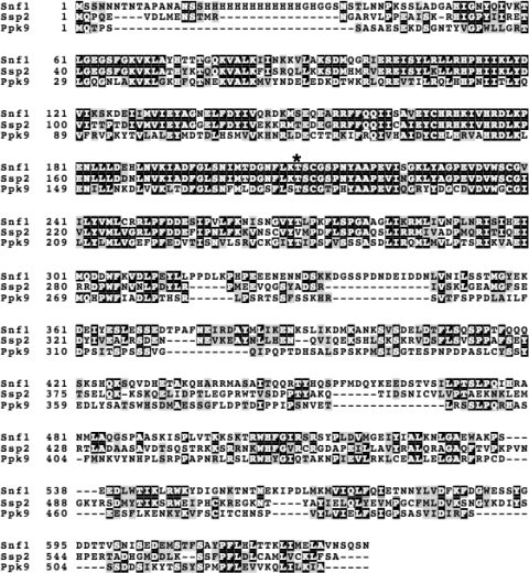
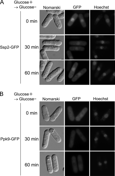
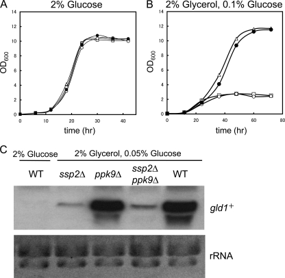
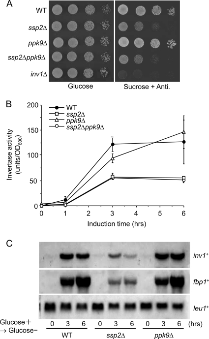
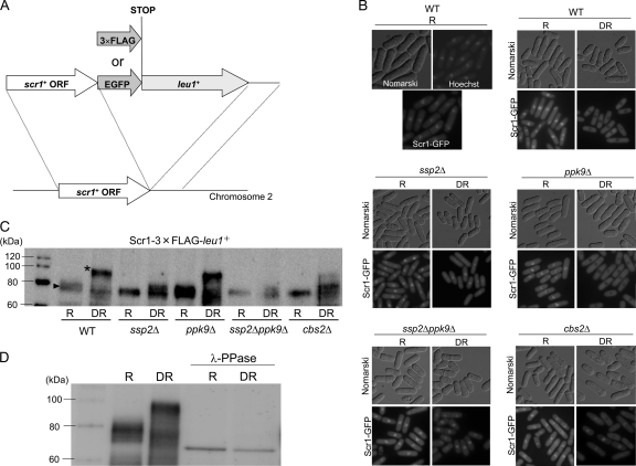
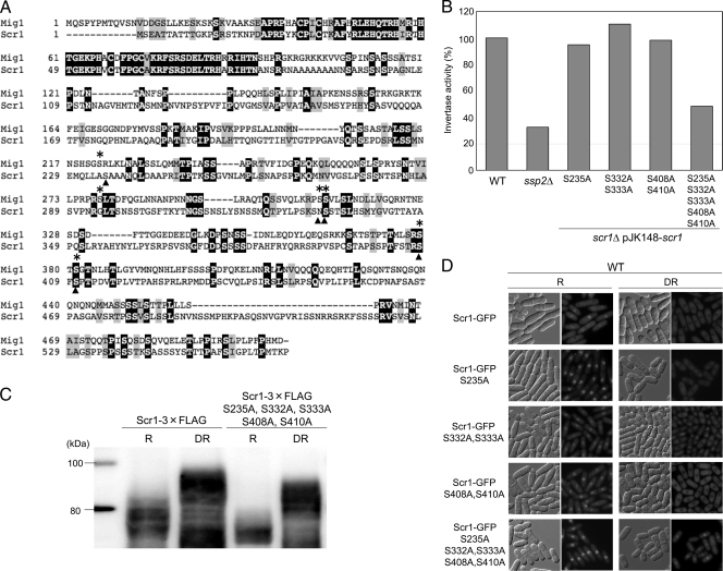
The function of two fission yeast genes, SPCC74.03c/ssp2(+) and SPAC23H4.02/ppk9(+), encoding an Snf1-like protein kinase were investigated. Deletion of ssp2(+) caused a partial defect in glucose derepression of inv1(+), fbp1(+), and gld1(+) and in assimilation of sucrose and glycerol, while a mutation in ppk9(+) had no apparent effect. Scr1, a transcription factor involved in glucose repression, localized to the nucleus under glucose-rich conditions and to the cytoplasm during glucose starvation in wild-type cells. In contrast, in the ssp2Δ mutant, Scr1 localized to the nucleus in cells grown in glucose-rich medium as well as in glucose-starved cells. Immunoblot analysis showed that Ssp2 is required for the phosphorylation of Scr1 upon glucose deprivation. Mutation of five putative Ssp2 recognition sites in Scr1 prevented glucose derepression of invertase in glucose-starved cells. These results indicate that Ssp2 regulates phosphorylation and subcellular localization of Scr1 in response to glucose.


Figures

Fig 1
Fig 1
Alignment of S. cerevisiae Snf1, S. pombe Ssp2, and S. pombe Ppk9. Amino acid residues identical to those in Snf1 are shaded. The threonine residue essential for activation of the Snf1 kinase (210T) is indicated with an asterisk.
Fig 2
Fig 2
Localization of Ssp2 and Ppk9. (A) Wild-type cells expressing Ssp2-GFP were cultured on MM containing 8% glucose and were harvested during logarithmic growth (0 min). Cells were then shifted to MM containing 2% glycerol and 0.05% glucose as carbon sources and incubated for 30 or 60 min. (B) Wild-type cells expressing Ppk9-GFP were cultured on MM containing 8% glucose and were harvested during logarithmic growth (0 min). Cells were then shifted to MM containing 2% glycerol and 0.05% glucose as carbon sources and incubated for 30 or 60 min. Nomarski differential interference contrast micrographs (Nomarski), GFP fluorescence (GFP), and Hoechst 33324 staining (Hoechst) are shown.
Fig 3
Fig 3
Glycerol assimilation and expression of gld1+ in ssp2Δ and ppk9Δ mutants. Wild-type (WT; filled circles), ssp2Δ (open circles), ppk9Δ (open triangles), and ssp2Δ ppk9Δ (open squares) strains were cultured on MM containing 2% glucose (A) and 2% glycerol and 0.1% glucose (B) as carbon sources. (C) Northern blot analysis of total RNA extracted from wild-type, ssp2Δ, ppk9Δ, and ssp2Δ ppk9Δ strains grown in MM containing 2% glucose or 2% glycerol and 0.05% glucose and harvested at an OD600 of 0.8.
Fig 4
Fig 4
Glucose derepression of invertase in ssp2Δ and ppk9Δ mutants. (A) Response of ssp2Δ and ppk9Δ mutants to sucrose availability. Identical volumes of 10-fold serial dilutions of exponentially growing wild-type parent strain, ssp2Δ, ppk9Δ, ssp2Δ ppk9Δ, and inv1Δ cells were spotted onto YES (left) or antimycin (Anti.; 10 μg/ml)-supplemented YES-sucrose medium (right) and incubated for 72 h at 30°C. (B) Glucose-derepression profiles of invertase in ssp2Δ and ppk9Δ mutants. Activity of secreted invertase in each strain was assayed at 0, 1, 3, and 6 h after shifting to medium containing 2% glycerol and 0.05% glucose as carbon sources. One unit of invertase activity is defined as the amount of enzyme which catalyzes production of 1 nmol of glucose per minute. Values are means ± SEMs (n = 3). (C) Northern blot analysis of inv1+ and fbp1+ in ssp2Δ and ppk9Δ mutants. Total RNA was harvested at 0, 3, and 6 h after glucose starvation (2% glycerol and 0.05% glucose) and subjected to analysis. Expression of leu1+ was determined as an internal control.
Fig 5
Fig 5
Localization and phosphorylation of Scr1. (A) Constructs expressing Scr1-GFP and Scr1-3×FLAG. Scr1-GFP and Scr1-3×FLAG were integrated at the scr1+ locus using leu1+ as a selectable marker. EGFP, enhanced GFP. (B) Localization of Scr1-GFP in wild-type, ssp2Δ, ppk9Δ, ssp2Δ ppk9Δ, and cbs2Δ strains. Scr1-GFP-expressing cells were cultured on MM containing 8% glucose to an OD600 of 0.8 (R). Cells were then shifted to MM containing 2% glycerol and 0.05% glucose as carbon sources and incubated for 1 h (DR). Nomarski differential interference contrast micrographs (Nomarski), GFP fluorescence (GFP), and Hoechst 33324 staining (Hoechst) are shown. (C) Immunoblot analysis of the Scr1-3×FLAG protein. Protein extracts were prepared from wild-type, ssp2Δ, ppk9Δ, ssp2Δ ppk9Δ, and cbs2Δ strains expressing Scr1-3×FLAG grown in MM containing 8% glucose (R) or 2% glycerol and 0.05% glucose (DR) and subjected to SDS-PAGE. (D) Protein extracts prepared from wild-type cells grown in MM medium containing 8% glucose (R) or 2% glycerol and 0.05% glucose (DR) were preincubated with phage lambda protein phosphatase (λ-PPase).
Fig 6
Fig 6
Effect of point mutations in Scr1 on glucose derepression of invertase, phosphorylation, and localization. (A) Alignment of S. cerevisiae Mig1 and S. pombe Scr1. Amino acid residues identical to those in Mig1 are shaded. Potential phosphorylation sites in Mig1 are indicated with an asterisk, and mutated sites in Scr1 are indicated with an arrowhead. (B) Glucose-derepression profiles of invertase in wild-type, ssp2Δ, and scr1Δ strains carrying a point-mutated scr1 gene. Strains were cultured in MM containing 8% glucose and shifted to MM containing 2% glycerol and 0.05% glucose as carbon sources and incubated for 3 h. Invertase activity of wild-type cells cultured under derepressing conditions was taken as 100%. Values are means ± SEMs (n = 3). (C) Immunoblot analysis of mutant Scr1-3×FLAG proteins. Wild-type cells expressing Scr1-3×FLAG or mutant Scr1-3×FLAG were cultured under glucose-repressing (R; 8% glucose) or glucose-derepressing (DR; 2% glycerol and 0.05% glucose) conditions. Proteins were extracted and subjected to SDS-PAGE. (D) Localization of mutant Scr1-GFP. Wild-type cells expressing Scr1-GFP or mutated Scr1-GFP were grown in MM containing 8% glucose to an OD600 of 0.8 (R). Cells were then shifted to MM containing 2% glycerol and 0.05% glucose as carbon sources and incubated for 1 h (DR). Nomarski differential interference contrast micrographs (Nomarski) and GFP fluorescence are shown.

Similar articles

Cited by

References

    1. Bimbo A, et al. 2005. Systematic deletion analysis of fission yeast protein kinases. Eukaryot. Cell 4:799–813 - PMC - PubMed
    1. Byrne SM, Hoffman CS. 1993. Six git genes encode a glucose-induced adenylate cyclase activation pathway in the fission yeast Schizosaccharomyces pombe. J. Cell Sci. 105:1095–1100 - PMC - PubMed
    1. Carlson M. 1999. Glucose repression in yeast. Curr. Opin. Microbiol. 2:202–207 - PubMed
    1. Carlson M, Osmond BC, Botstein D. 1981. Mutants of yeast defective in sucrose utilization. Genetics 98:25–40 - PMC - PubMed
    1. Celenza JL, Eng FJ, Carlson M. 1989. Molecular analysis of the SNF4 gene of Saccharomyces cerevisiae: evidence for physical association of the SNF4 protein with the SNF1 protein kinase. Mol. Cell. Biol. 9:5045–5054 - PMC - PubMed

Publication types

MeSH terms

Substances