

Adipose tissue stem cells meet preadipocyte commitment: going back to the future
- PMID: 22140268
- PMCID: PMC3269153
- DOI: 10.1194/jlr.R021089
Adipose tissue stem cells meet preadipocyte commitment: going back to the future
Abstract
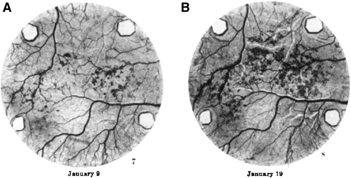
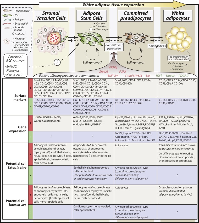
White adipose tissue (WAT) is perhaps the most plastic organ in the body, capable of regeneration following surgical removal and massive expansion or contraction in response to altered energy balance. Research conducted for over 70 years has investigated adipose tissue plasticity on a cellular level, spurred on by the increasing burden that obesity and associated diseases are placing on public health globally. This work has identified committed preadipocytes in the stromal vascular fraction of adipose tissue and led to our current understanding that adipogenesis is important not only for WAT expansion, but also for maintenance of adipocyte numbers under normal metabolic states. At the turn of the millenium, studies investigating preadipocyte differentiation collided with developments in stem cell research, leading to the discovery of multipotent stem cells within WAT. Such adipose tissue-derived stem cells (ASCs) are capable of differentiating into numerous cell types of both mesodermal and nonmesodermal origin, leading to their extensive investigation from a therapeutic and tissue engineering perspective. However, the insights gained through studying ASCs have also contributed to more-recent progress in attempts to better characterize committed preadipocytes in adipose tissue. Thus, ASC research has gone back to its roots, thereby expanding our knowledge of preadipocyte commitment and adipose tissue biology.
Figures
References
-
- Fleck S. J. 1983. Body composition of elite American athletes. Am. J. Sports Med. 11: 398–403. - PubMed
-
- Ortega F. J., Mayas D., Moreno-Navarrete J. M., Catalan V., Gomez-Ambrosi J., Esteve E., Rodriguez-Hermosa J. I., Ruiz B., Ricart W., Peral B., et al. 2010. The gene expression of the main lipogenic enzymes is downregulated in visceral adipose tissue of obese subjects. Obesity (Silver Spring). 18: 13–20. - PubMed
-
- Larson K. A., Anderson D. B. 1978. The effects of lipectomy on remaining adipose tissue depots in the Sprague Dawley rat. Growth. 42: 469–477. - PubMed
-
- Reyne Y., Nougues J., Vezinhet A. 1983. Adipose tissue regeneration in 6-month-old and adult rabbits following lipectomy. Proc. Soc. Exp. Biol. Med. 174: 258–264. - PubMed
-
- Hernandez T. L., Kittelson J. M., Law C. K., Ketch L. L., Stob N. R., Lindstrom R. C., Scherzinger A., Stamm E. R., Eckel R. H. 2011. Fat redistribution following suction lipectomy: defense of body fat and patterns of restoration. Obesity (Silver Spring). 19: 1388–1395. - PubMed
Publication types
MeSH terms
Substances
Grants and funding
LinkOut - more resources
Full Text Sources
Other Literature Sources
Miscellaneous
