. 2012 Jan;49(1):58-65.
doi: 10.1136/jmedgenet-2011-100174. Epub 2011 Dec 3.

A genome-wide association study of men with symptoms of testicular dysgenesis syndrome and its network biology interpretation

Affiliations

A genome-wide association study of men with symptoms of testicular dysgenesis syndrome and its network biology interpretation

Marlene D Dalgaard et al. J Med Genet. 2012 Jan.

Abstract

Background: Testicular dysgenesis syndrome (TDS) is a common disease that links testicular germ cell cancer, cryptorchidism and some cases of hypospadias and male infertility with impaired development of the testis. The incidence of these disorders has increased over the last few decades, and testicular cancer now affects 1% of the Danish and Norwegian male population.

Methods: To identify genetic variants that span the four TDS phenotypes, the authors performed a genome-wide association study (GWAS) using Affymetrix Human SNP Array 6.0 to screen 488 patients with symptoms of TDS and 439 selected controls with excellent reproductive health. Furthermore, they developed a novel integrative method that combines GWAS data with other TDS-relevant data types and identified additional TDS markers. The most significant findings were replicated in an independent cohort of 671 Nordic men.

Results: Markers located in the region of TGFBR3 and BMP7 showed association with all TDS phenotypes in both the discovery and replication cohorts. An immunohistochemistry investigation confirmed the presence of transforming growth factor β receptor type III (TGFBR3) in peritubular and Leydig cells, in both fetal and adult testis. Single-nucleotide polymorphisms in the KITLG gene showed significant associations, but only with testicular cancer.

Conclusions: The association of single-nucleotide polymorphisms in the TGFBR3 and BMP7 genes, which belong to the transforming growth factor β signalling pathway, suggests a role for this pathway in the pathogenesis of TDS. Integrating data from multiple layers can highlight findings in GWAS that are biologically relevant despite having border significance at currently accepted statistical levels.


Conflict of interest statement

Competing interests: None.

Figures

Figure 1
Figure 1
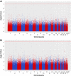
Manhattan plots showing the association between all single-nucleotide polymorphisms and (A) testicular dysgenesis syndrome and (B) the subset of cases of testicular germ cell cancer.
Figure 2
Figure 2
Integration of genome-wide association study (GWAS) data with heterogeneous data types. The rationale behind the method is to prioritise genes if several data types show mildly pronounced associations with the phenotype, thereby identifying genes that would not have been found with GWAS analysis alone. TGFBR3, which was selected by this method and whose single-nucleotide polymorphism markers were further validated, is shown in red. The position of TGFBR3 is marked by red circles in the distributions of p values, and by red squares in the ranking of each data type layer. Three data types were used: (1) single-marker GWAS results of this study (grey); (2) targeted mutations from the Mouse Genome Informatics (MGI) database, filtered for testicular dysgenesis syndrome phenotypes, and ranked by their enrichment in protein–protein complexes (pink); (3) differential expression in the fetal testis of mouse and human (green). (A) A distribution of the p values of all human genes is shown for each of the data types. The p value ranges from 0 to 1, with 0 at the top. The p value associated with each gene is converted into a rank, such that each human gene is assigned a rank. The vertical line to the right of each distribution represents the ranking of all genes, where the top corresponds to rank 1. (B) The gene ranks of each individual data type are combined to a final meta-rank of each gene. As an example, TGFBR3 had the ranks 238, 42 and 408 among all human genes in the three data types: GWAS, targeted mutations with protein–protein interaction enrichment, and differential expression in the developing testis, respectively. After combination of these data type-specific ranks, TGFBR3 was ranked 3rd among all genes. PPI, protein-protein interaction.
Figure 3
Figure 3
Regional association plots and linkage disequilibrium structure. The −log10 of the p values for the association of discovery markers are shown, and the markers are coloured in a white to red scale according to the strength of the pairwise linkage disequilibrium (r2) to the most significant discovery marker at each locus. The blue marker represents the association in the replication stage. Light blue peaks indicate recombination rates from the CEU HapMap population. r2-based linkage disequilibrium structures from the genome-wide association study data are displayed at the bottom. (A) The strongest association in the discovery stage was found at 2q31.1, close to the cluster of HOXD genes. (B) The most significant markers in the replication stage were found at the KITLG region. The genes TGFBR3 (C) and PBM7 (D) had single-nucleotide polymorphisms with mild association in the discovery stage, but were top-ranked by the integrative data analysis, and were further validated in the replication stage.
Figure 4
Figure 4
Localisation of transforming growth factor β receptor type III (TGFBR3) protein in normal fetal testis (A), adult testis with complete spermatogenesis (B), and dysgenetic adult testis with carcinoma in situ and Leydig cell hyperplasia (C). Leydig cells are marked by arrowheads (A, B, C), gonocytes (A) and CIS cells (C) by large arrows and peritubular cells by small arrows (B & C). Inserts (lower right) are without primary antibody. Bars indicate 100 μm. TGFBR3 is seen to be expressed in Leydig and peritubular cells.

References

    1. Schnack TH, Poulsen G, Myrup C, Wohlfahrt J, Melbye M. Familial coaggregation of cryptorchidism, hypospadias, and testicular germ cell cancer: a nationwide cohort study. J Natl Cancer Inst 2010;102:187–92 - PubMed
    1. Skakkebaek NE, Rajpert-De Meyts E, Main KM. Testicular dysgenesis syndrome: an increasingly common developmental disorder with environmental aspects. Hum Reprod 2001;16:972–8 - PubMed
    1. Purdue MP, Devesa SS, Sigurdson AJ, McGlynn KA. International patterns and trends in testis cancer incidence. Int J Cancer 2005;115:822–7 - PubMed
    1. Engholm G, Ferlay J, Christensen N, Bray F, Gjerstorff ML, Klint A, Køtlum JE, Olafsdóttir E, Pukkala E, Storm HH. NORDCAN–a Nordic tool for cancer information, planning, quality control and research. Acta Oncol 2010;49:725–36 - PubMed
    1. Asklund C, Jensen TK, Main KM, Sobotka T, Skakkebaek NE, Jørgensen N. Semen quality, reproductive hormones and fertility of men operated for hypospadias. Int J Androl 2010;33:80–7 - PubMed

Publication types

MeSH terms