

A new look at glutamate and ischemia: NMDA agonist improves long-term functional outcome in a rat model of stroke
- PMID: 22140354
- PMCID: PMC3229223
- DOI: 10.2217/fnl.11.55
A new look at glutamate and ischemia: NMDA agonist improves long-term functional outcome in a rat model of stroke
Abstract
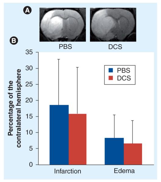
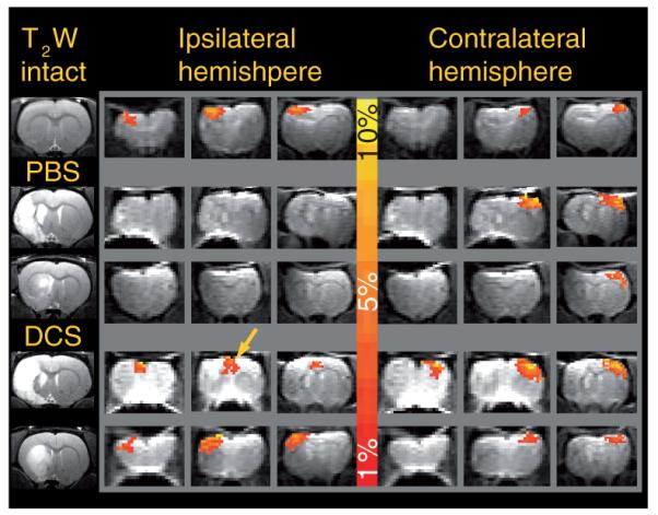
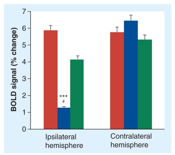
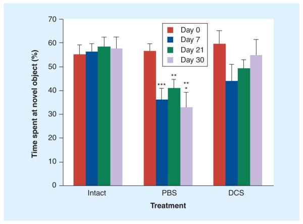
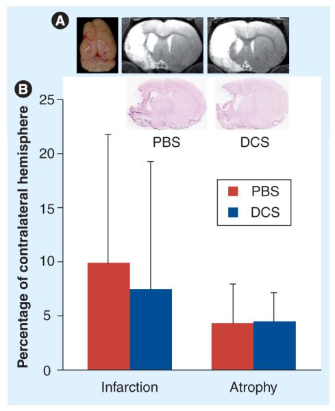
Ischemic stroke triggers a massive, although transient, glutamate efflux and excessive activation of NMDA receptors (NMDARs), possibly leading to neuronal death. However, multiple clinical trials with NMDA antagonists failed to improve, or even worsened, stroke outcome. Recent findings of a persistent post-stroke decline in NMDAR density, which plays a pivotal role in plasticity and memory formation, suggest that NMDAR stimulation, rather than inhibition, may prove beneficial in the subacute period after stroke. AIM: This study aims to examine the effect of the NMDAR partial agonist d-cycloserine (DCS) on long-term structural, functional and behavioral outcomes in rats subjected to transient middle cerebral artery occlusion, an animal model of ischemic stroke. MATERIALS #ENTITYSTARTX00026; METHODS: Rats (n = 36) that were subjected to 90 min of middle cerebral artery occlusion were given a single injection of DCS (10 mg/kg) or vehicle (phosphate-buffered saline) 24 h after occlusion and followed up for 30 days. MRI (structural and functional) was used to measure infarction, atrophy and cortical activation due to electrical forepaw stimulation. Memory function was assessed on days 7, 21 and 30 postocclusion using the novel object recognition test. A total of 20 nonischemic controls were included for comparison. RESULTS: DCS treatment resulted in significant improvement of somatosensory and cognitive function relative to vehicle treatment. By day 30, cognitive performance of the DCS-treated animals was indistinguishable from nonischemic controls, while vehicle-treated animals demonstrated a stable memory deficit. DCS had no significant effect on infarction or atrophy. CONCLUSION: These results support a beneficial role for NMDAR stimulation during the recovery period after stroke, most likely due to enhanced neuroplasticity rather than neuroprotection.
Figures
Similar articles
-
Novel approach to the role of NMDA receptors in traumatic brain injury.CNS Neurol Disord Drug Targets. 2014;13(4):567-73. doi: 10.2174/18715273113126660196. CNS Neurol Disord Drug Targets. 2014. PMID: 24168367 Review.
-
Melatonin improves neuroplasticity by upregulating the growth-associated protein-43 (GAP-43) and NMDAR postsynaptic density-95 (PSD-95) proteins in cultured neurons exposed to glutamate excitotoxicity and in rats subjected to transient focal cerebral ischemia even during a long-term recovery period.J Pineal Res. 2014 Mar;56(2):213-23. doi: 10.1111/jpi.12114. Epub 2014 Jan 13. J Pineal Res. 2014. PMID: 24350898
-
Lithium upregulates growth-associated protein-43 (GAP-43) and postsynaptic density-95 (PSD-95) in cultured neurons exposed to oxygen-glucose deprivation and improves electrophysiological outcomes in rats subjected to transient focal cerebral ischemia following a long-term recovery period.Neurol Res. 2022 Oct;44(10):870-878. doi: 10.1080/01616412.2022.2056817. Epub 2022 Mar 29. Neurol Res. 2022. PMID: 35348035
-
GLYX-13, a NMDA Receptor Glycine-Site Functional Partial Agonist, Attenuates Cerebral Ischemia Injury In Vivo and Vitro by Differential Modulations of NMDA Receptors Subunit Components at Different Post-Ischemia Stage in Mice.Front Aging Neurosci. 2017 Jun 9;9:186. doi: 10.3389/fnagi.2017.00186. eCollection 2017. Front Aging Neurosci. 2017. PMID: 28649199 Free PMC article.
-
D-cycloserine in Schizophrenia: New Strategies for Improving Clinical Outcomes by Enhancing Plasticity.Curr Neuropharmacol. 2017;15(1):21-34. doi: 10.2174/1570159x14666160225154812. Curr Neuropharmacol. 2017. PMID: 26915421 Free PMC article. Review.
Cited by
-
Region-specific role for GluN2B-containing NMDA receptors in injury to Purkinje cells and CA1 neurons following global cerebral ischemia.Neuroscience. 2015 Jan 22;284:555-565. doi: 10.1016/j.neuroscience.2014.10.033. Epub 2014 Oct 24. Neuroscience. 2015. PMID: 25450957 Free PMC article.
-
Neuroprotective Effect of ZnT3 Knockout on Subarachnoid Hemorrhage.Transl Neurosci. 2018 Jul 4;9:26-32. doi: 10.1515/tnsci-2018-0006. eCollection 2018. Transl Neurosci. 2018. PMID: 29992050 Free PMC article.
-
Protection from cerebral ischemia by inhibition of TGFβ-activated kinase.Exp Neurol. 2012 Sep;237(1):238-45. doi: 10.1016/j.expneurol.2012.05.019. Epub 2012 Jun 5. Exp Neurol. 2012. PMID: 22683931 Free PMC article.
-
Neuropathophysiology of Brain Injury.Anesthesiol Clin. 2016 Sep;34(3):453-64. doi: 10.1016/j.anclin.2016.04.011. Anesthesiol Clin. 2016. PMID: 27521191 Free PMC article. Review.
-
NAAG peptidase inhibitors and deletion of NAAG peptidase gene enhance memory in novel object recognition test.Eur J Pharmacol. 2013 Feb 15;701(1-3):27-32. doi: 10.1016/j.ejphar.2012.11.027. Epub 2012 Nov 29. Eur J Pharmacol. 2013. PMID: 23200894 Free PMC article.
References
-
- Carandang R, Seshadri S, Beiser A. Trends in incidence, lifetime risk, severity, and 30-day mortality of stroke over the past 50 years. JAMA. 2006;296:2939–2946. - PubMed
-
- Rosamond W, Flegal K, Furie K, et al. Heart disease and stroke statistics 2008 update a report from the American Heart Association Statistics Committee and Stroke Statistics Subcommittee. Circulation. 2008;117:E25–E146. - PubMed
-
- Wang KM, Kyoung KP, Yong SK, et al. Atherothrombotic middle cerebral artery territory infarction: topographic diversity with common occurrence of concomitant small cortical and subcortical infarcts. Stroke. 2000;31:2055–2061. - PubMed
-
- Caplan L, Schmahmann J, Kase C, et al. Caudate infarcts. Arch. Neurol. 1990;47:133–143. - PubMed
-
- Phipps L. Assessment of neurologic deficits in stroke: acute-care and rehabilitation implications. Nurs. Clin. North Am. 1991;26:957–970. - PubMed
Grants and funding
LinkOut - more resources
Full Text Sources