. 2011;6(11):e27438.
doi: 10.1371/journal.pone.0027438. Epub 2011 Nov 30.

A systematic analysis of the 3'UTR of HNF4A mRNA reveals an interplay of regulatory elements including miRNA target sites

Affiliations

A systematic analysis of the 3'UTR of HNF4A mRNA reveals an interplay of regulatory elements including miRNA target sites

Andrea Wirsing et al. PLoS One. 2011.

Abstract

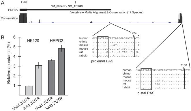
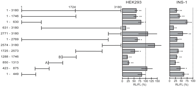
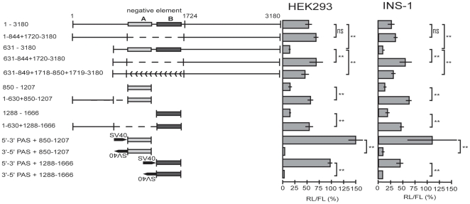
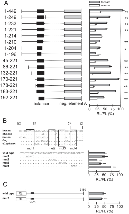
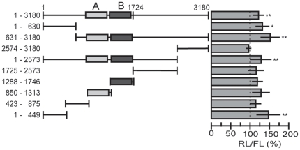
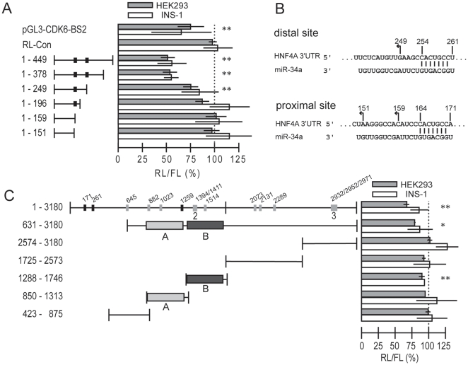
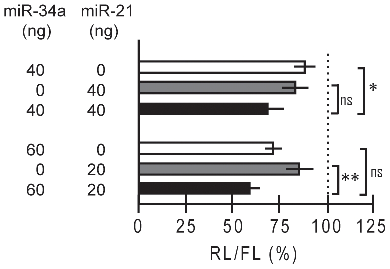
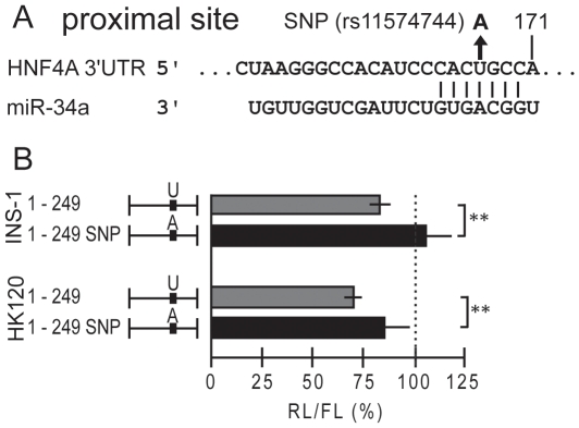
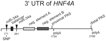
Dysfunction of hepatocyte nuclear factor 4α (HNF4α) has been linked to maturity onset diabetes of the young (MODY1), diabetes type II and possibly to renal cell carcinoma (RCC). Whereas diabetes causing mutations are well known, there are no HNF4A mutations found in RCC. Since so far analyses have been constricted to the promoter and open reading frame of HNF4A, we performed a systematic analysis of the human HNF4A 3'UTR. We identified a short (1724 nt) and long (3180 nt) 3'UTR that are much longer than the open reading frame and conferred a repressive effect in luciferase reporter assays in HEK293 and INS-1 cells. By dissecting the 3'UTR into several pieces, we located two distinct elements of about 400 nt conferring a highly repressive effect. These negative elements A and B are counteracted by a balancer element of 39 nt located within the 5' end of the HNF4A 3'UTR. Dicer knock-down experiments implied that the HNF4A 3'UTR is regulated by miRNAs. More detailed analysis showed that miR-34a and miR-21 both overexpressed in RCC cooperate in downregulation of the HNF4A mRNA. One of the identified miR-34a binding sites is destroyed by SNP rs11574744. The identification of several regulatory elements within the HNF4A 3'UTR justifies the analysis of the 3'UTR sequence to explore the dysfunction of HNF4α in diabetes and RCC.


Conflict of interest statement

Competing Interests: The authors have declared that no competing interests exist.

Figures

Figure 1
Figure 1. Two distinct polyadenylation signals (PAS) in the human HNF4A mRNA.
(A) Schematic representation of the HNF4A 3′UTR. The screen shot taken from the UCSC Genome Browser (assembly March 2006) depicts the known human HNF4A 3′UTR with the RefSeq sequences NM_000457.3 and NM_178849.1 and the genome position from 42,491,540 to 42,494,950 of chromosome 20. The degree of conservation across 17 species is indicated by black areas. The nucleotide sequence alignment of the region surrounding the proximal and distal PAS is shown below. Non-conserved nucleotides in comparison to the human sequence are given for the different species, while dots represent conserved nucleotides. The proximal and distal PAS are boxed and the corresponding cleavage sites, as determined by 3′ RACE and subsequent sequencing, are indicated by a vertical line. The last nucleotide of the short and long 3′UTR is marked at position 1724 and 3180, respectively. (B) The relative abundance of the short and long HNF4A 3′UTRs was determined in comparison to the house keeping gene GAPDH. Two independent RNA samples were prepared from each cell line and the qRT-PCR was performed in triplicates. Each column thus represents the mean±SD of six measurements.
Figure 2
Figure 2. Systematic reporter analyses of the human HNF4A 3′UTR.
The results of luciferase assays 24 h after transient transfection into HEK293 and INS-1 cells are shown. The numbers of the construct names refer to the nucleotide position in the HNF4A 3′UTR with 1 being the first nucleotide after the stop codon. Each 3′UTR fragment was cloned downstream of the Renilla luciferase ORF into the RL-Con plasmid. At least three transfection assays were performed for each construct, involving two independent plasmid preparations. Each assay was performed in triplicate and as indicated by RL/FL a CMV-driven firefly luciferase (FL) was used to control for transfection efficiency. The activity of the empty RL-Con plasmid was used for standardization to 100%. p-values were determined using a one-sample t test. p-values of <0.05 and of <0.01 are indicated by * or **, respectively.
Figure 3
Figure 3. Balancer counteracting the negative elements A and B.
The negative elements A and B are indicated by light grey and dark grey boxes, respectively. The deletion of the negative elements is illustrated by a broken line, whereas the inversion of this element is marked by backwards arrows. The insertion of the SV40 termination signal in sense and antisense is marked. The results of luciferase assays were derived and evaluated as in Fig. 2. p-values were determined between two columns as indicated by brackets using an independent-samples t test. Non-significant changes are marked by ns and refer to p-values>0.05 and ** refers to p-values<0.01.
Figure 4
Figure 4. Mapping of the balancer element counteracting the negative elements.
(A) The indicated 5′ sequences of the HNF4A 3′UTR were cloned upstream of negative element A (850–1207, light grey box). The identified balancer is marked by a black box. Luciferase assays were performed in INS-1 cells as described for Fig. 2. Grey and white bars represent results obtained with the constructs containing the 5′ sequences in front of negative element A in forward (5′-3′) and reverse orientation (3′-5′), respectively. The results of luciferase assays were derived and evaluated as in Fig. 2. p-values of<0.01 and of <0.001, determined between the forward and reverse orientation of each construct, are indicated by * or **, respectively. (B) The balancer sequence identified in the human 3′UTR of HNF4A was aligned to the corresponding sequence of other mammals using the UCSC Genome Browser (hg18, multiz alignments of 44 vertebrates). The four regions conserved are boxed. Below, the wild type balancer sequence and the four mutants tested in front of the negative element A (850–1207) are given. Their performance in the luciferase assay in INS-1 cells is summarized from at least three independent preparation of each construct. p-values of<10−5 (***) are calculated in comparison to the wild type sequence using an independent-samples t test. (C) The long (1–3180) 3′UTR linked downstream of the Renilla luciferase (RL) and a corresponding construct containing mut2 given in panel B were assayed in INS-1 cells. The balancer (grey box) and mut2 (black box) are not drawn to scale. Eight and ten independent plasmid preparations were used for the wild type and mut2 construct, respectively. p-values of<10−5 (***) are calculated in comparison to the wild type sequence using an independent-samples t test.
Figure 5
Figure 5. Dicer knock-down indicates that the HNF4A 3′UTR is regulated by miRNAs.
To knock-down the Dicer protein, doxycycline (1 µg/ml) was added to the Dicer-kd/2b2 cell line for three or seven days. Two days before luciferase activity was measured, the cells were transiently transfected with reporter constructs. The nomenclature of the constructs is as in Fig. 2. At least three transfection assays were performed for each construct, involving at least two independent plasmid preparations. Each assay was performed in triplicate and a CMV-driven firefly luciferase (FL) was used to control for transfection efficiency. The activity of each construct measured in the presence of Dicer (ethanol added) was used for standardization (100%) and is not shown. The 3xBulgeB and RL-Con reporter plasmids harboring three bulged binding sites for let-7a and lacking any binding sites, respectively, were included as a positive and negative control in each experiment (not shown). The negative elements A and B identified in Fig. S1 are indicated. The p-values were determined using an independent-samples t test. p-values of<0.05 and of <0.01 are indicated by * or **, respectively.
Figure 6
Figure 6. Reporter analyses of miR-34a binding sites in the HNF4A 3′UTR.
(A) miR-34a targets two sites within the 5′ 449 nt of the HNF4A 3′UTR. Reporter plasmids and pri-miR-34a expression plasmids were co-transfected 24 h before cell collection, into HEK293 (upper grey bars) and INS-1 cells (lower white bars). At least one transfection assay was performed for each construct, involving two independent plasmid preparations in the case of two or more assays. Each assay was performed in triplicate and a CMV-driven firefly luciferase was used to control for transfection efficiency. The activity of each construct in the absence of the pri-miR-34a plasmid (replaced by Rc/CMV) was used for standardization (100%) and is not shown. pGL3-CDK6-BS2 and pRL-Con served as positive and negative controls, respectively. Since pGL3-CDK6-BS2 expresses the firefly luciferase, the RL-Con plasmid was used to control for transfection efficiency. The black boxes indicate miR-34a target sites with perfect seed sequence. To calculate p-values the data of HEK293 and INS-1 cells were combined for each construct. p-values are<0.05 (*) and <0.01 (**) using an independent-samples t test. (B) Schematic diagram of the two potential miR-34a binding sites within the 5′ 449 nt of the HNF4A 3′UTR. The numbering refers to the first nucleotide after the stop codon as 1. The site extending from 149–171 nt was only predicted by TargetScan and is little conserved, while the distal site located at 239–261 nt was predicted by RNA22 and TargetScan and is highly conserved among vertebrates. The two perfect seed matches are indicated by vertical lines between the HNF4A 3′UTR and miR-34a sequence. The arrows at position 151, 159 and 249 mark the last nucleotide of the HNF4A 3′UTR in the corresponding constructs. (C) Analyzing the performance of various 3′UTR fragments upon miR-34a overexpression as performed in panel A. The grey and black boxes indicate miR-34a target sites without and with a perfect seed sequence (CACUGCC), respectively, as determined by RNA22 . The number of target sites indicated by a box is given underneath the target site in case of more than one site. The nucleotide positions of the 3′ end of the target sites are given. The negative elements A and B identified in Fig. S1 are indicated.
Figure 7
Figure 7. miR-34a and miR-21 cooperate on the HNF4A 3′UTR.
The reporter plasmid with the full-length HNF4A 3′UTR (1–3180) was cotransfected into INS-1 cells with expression vectors encoding pri-miR-34a and/or a miR-21 as indicated. 24 h later Renilla luciferase activity was measured and standardized using the cotransfected firefly luciferase reporter. Each experiment involved three assays performed in triplicate and the activity in the presence of the empty expression vector was used for standardization (100%). By adding empty vector the amount of DNA was kept constant. The specificity of the miR-21 expression vector pCMV-miR21 was verified by using the bona fide reporter Luc-TPM1-V1-UTR and its derivative Luc-TPM1-V4-UTR lacking a miR-21 binding site (Fig. S2). p-values of >0.05 (ns), <0.05 (*) and <0.01 (**) are calculated as indicated by brackets using an independent-samples t test.
Figure 8
Figure 8. SNP rs11574744 destroys the proximal miR-34a binding site.
(A) The nucleotide change of the SNP rs11574744 in the proximal miR-34a site 171 nt downstream of the stop codon of the HNF4A mRNA is given. (B) Luciferase reporter assays were performed in INS-1 and HK120 cells with the expression vector for miR-34a and either the HNF4A 3′UTR reporter construct 1–249 or the corresponding construct carrying the A nucleotide variant. 100% refers to the activity in the presence of an empty expression vector. ** denotes p-values of <0.01 using an independent-samples t test. Experimental details are as in Fig. 6.
Figure 9
Figure 9. Regulatory elements identified in the 3′UTR of the HNF4A mRNA.
A summary of the various functional elements identified in our work is illustrated. The SNP rs11574744 located in the proximal miR-34a site (171 nt) is given. The functional relevance of the distal miR-34a site (271 nt) has been reported independently . The numbering starts with the first nucleotide of the 3′UTR after the stop codon.

Similar articles

Cited by

References

    1. Bolotin E, Schnabl JM, Sladek FM. HNF4A (Homo sapiens). Transcription Factor Encyclopedia. 2010. Available: http://www.cisreg.ca/tfe. Accessed 2010 July 15.
    1. Bolotin E, Sladek FM, Schnabl JM. Hnf4a (Mus musculus). Transcription Factor Encyclopedia. 2010. Available: http://www.cisreg.ca/tfe. Accessed 2010 July 15.
    1. Odom DT, Zizlsperger N, Gordon DB, Bell GW, Rinaldi NJ, et al. Control of pancreas and liver gene expression by HNF transcription factors. Science. 2004;303:1378–1381. - PMC - PubMed
    1. Erdmann S, Senkel S, Arndt T, Lucas B, Lausen J, et al. Tissue-specific transcription factor HNF4alpha inhibits cell proliferation and induces apoptosis in the pancreatic INS-1 beta-cell line. Biol Chem. 2007;388:91–106. - PubMed
    1. Grigo K, Wirsing A, Lucas B, Klein-Hitpass L, Ryffel GU. HNF4alpha orchestrates a set of 14 genes to down-regulate cell proliferation in kidney cells. Biol Chem. 2008;389:179–187. - PubMed

Publication types

MeSH terms