. 2011;6(11):e27680.
doi: 10.1371/journal.pone.0027680. Epub 2011 Nov 29.

A quantitative method for the specific assessment of caspase-6 activity in cell culture

Affiliations

A quantitative method for the specific assessment of caspase-6 activity in cell culture

Dagmar E Ehrnhoefer et al. PLoS One. 2011.

Abstract

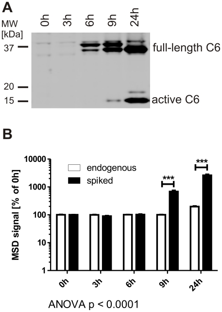
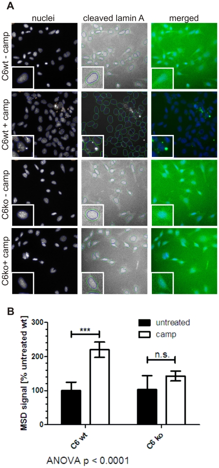
Aberrant activation of caspase-6 has recently emerged as a major contributor to the pathogeneses of neurodegenerative disorders such as Alzheimer's and Huntington disease. Commercially available assays to measure caspase-6 activity commonly use the VEID peptide as a substrate. However these methods are not well suited to specifically assess caspase-6 activity in the presence of other, confounding protease activities, as often encountered in cell and tissue samples. Here we report the development of a method that overcomes this limitation by using a protein substrate, lamin A, which is highly specific for caspase-6 cleavage at amino acid 230. Using a neo-epitope antibody against cleaved lamin A, we developed an electrochemiluminescence-based ELISA assay that is suitable to specifically detect and quantify caspase-6 activity in highly apoptotic cell extracts. The method is more sensitive than VEID-based assays and can be adapted to a high-content imaging platform for high-throughput screening. This method should be useful to screen for and characterize caspase-6 inhibitor compounds and other interventions to decrease intracellular caspase-6 activity for applications in neurodegenerative disorders.


Conflict of interest statement

Competing Interests: The Pfizer Innovation fund funded part of the work that was done at the Centre for Drug Research and Development (CDRD). The authors do not have any employment or consultant relationships with Pfizer, there are no relevant patents or products. The authors confirm that the funding does not alter their adherence to the PLos ONE policies on sharing data and materials.

Figures

Figure 1
Figure 1. The lamin A protein is a more specific substrate than its VEID peptide.
A: 100 µM VEID-Afc was incubated with different amounts of caspase -3, -6 or -7 for 1 h at 37°C. Fluorescence generated by cleavage was monitored over time, and the initial, linear portion of the curve was used to calculate the reaction velocity. VEID is preferentially cleaved by caspase-6, but also cross-reacts with caspases -3 and -7 at higher concentrations. Error bars are the SEM of N≥3 of 3 independent experiments. B: 5 µM VEID-Afc was incubated with 0.5 µM caspase-6 or different amounts of caspase-3 for 1 h at 37°C. The reaction velocity was calculated as in A. Even at this low concentration of VEID-Afc, the peptide substrate can be cleaved by caspase-3, and an 8 fold higher molar concentration of caspase-3 than caspase-6 results in a higher signal for VEID cleavage by caspase-3 than caspase-6. Error bars are the SEM of N = 3 independent experiments, statistical significance was assessed by 1-way ANOVA and post-hoc Dunnett comparisons: *** p<0.0001. C: Pure lamin A protein was incubated with different amounts of caspase -3, -6 or -7 for 30 min at 37°C. Samples were separated by SDS-PAGE and both fragments of cleaved lamin A was detected by Western blotting with antibodies #2031 (full-length lamin A and N-terminal fragment) and #2032 (C-terminal fragment). No cleavage was observed with caspases -3 or -7, while caspase-6 generated lamin A fragments in a dose-dependent manner. A representative image of 3 independent experiments is shown.
Figure 2
Figure 2. VEID, but not lamin A+C, is cleaved in the absence of caspase-6.
A: Caspase-6 protein (full-length, 32 kDa) is detected in MEFs generated from C6wt, but not C6ko mice. B: Endogenous lamin A protein (70 kDa) is cleaved in wt, but not C6ko MEFs after staurosporine stress for 4 h or longer. The antibody cross-reacts with full-length lamin C (60 kDa), and the cleaved band at 28 kDa has the same size for both lamin A+C (lower panel). C: MCF-7 cells express caspase-6, but not caspase-3 protein, whereas both C6wt and C6ko MEFs contain both caspases. hu: human, m: mouse, ns: non-specific band. D: MCF-7 cells were stressed with 5 µM camptothecin for different amounts of time and the cleavage of endogenous lamin A and C proteins was monitored by Western blotting with antibodies antibodies #2031 (full-length lamin A+C) and #2032 (C-terminal fragments). E: Schematic representation of lamin A and C and the caspase-6 cleavage site at AA 230. The N-terminal fragments generated by caspase-6 cleavage (red) have the same size (28 kDa) for both lamin A+C. F: C6wt or C6ko MEFs were stressed with 50 nM staurosporine for different amounts of time, lysates were generated and analyzed for cleavage of VEID-Afc. C6wt cells show a significant increase in fluorescence at each timepoint, the fluorescence signal obtained from C6ko lysates only reach a statistically significant difference from baseline after 4 h. C6ko MEFs stressed with staurosporine for 4 h or more show the same levels of VEID proteolysis as wt cells. Error bars are the SEM of N = 3 of 4 independent experiments. Statistical significance was assessed by 2-way ANOVA and post-hoc Bonferroni comparisons: *** p<0.0001, ** p<0.001, * p<0.01.
Figure 3
Figure 3. Increased sensitivity and highly improved specificity in a lamin-based caspase-6 activity assay.
A+B: Quantities of down to 10 nM active caspase-6 can be detected with the novel lamin A-based caspase-6 assay while maintaining a signal-noise ratio of >3. C: The peptide inhibitor VEID-CHO shows a similar IC50 value in both the VEID- and the lamin A- based caspase-6 assays. D: The lamin A-based caspase-6 assay is highly specific, with no signal generated from C6ko cells even after 6 h of staurosporine stress. Error bars are the SEM of N≥3 of 3 independent experiments. Statistical significance was assessed by 2-way ANOVA and post-hoc Bonferroni comparisons: *** p<0.0001, ** p<0.001.
Figure 4
Figure 4. Cleavage of lamin B1 in primary neuronal cultures is not caspase-6 specific, but spiking with lamin A provides a specific readout.
A: Lamins B1 and B2 differ from lamins A and C in their caspase cleavage site (VEVD versus VEID, respectively). B: Lamin B1 is cleaved after camptothecin stress in primary cortical neuronal cultures derived from C6wt and C6ko mice. C: Spiking with lamin A protein results in an increased ELISA signal in lysates from camptothecin-stressed C6wt, but not C6ko neurons. Boxes represent the 25th–75th percentile, whiskers represent the minimum and maximum. Data from 5 (C6ko) and 7 (C6wt) separate cultures are shown. Statistical significance was assessed by 2-way ANOVA and post-hoc Bonferroni comparisons: *** p<0.0001.
Figure 5
Figure 5. Spiking with lamin A allows for the detection of caspase-6 activity before the cleavage of endogenous lamins.
A: COS7 cells were transiently transfected with recombinant human caspase-6 and cultured for the indicated amounts of time. The active caspase-6 fragment can be observed by Western blotting 9 h after transfection and is increasingly abundant after 24 h. B: COS7 cells were transfected as in A. The cleavage of endogenous lamin A protein can be observed with the ELISA method after 24 h, whereas spiking of lysates with lamin A protein leads to a robust detection of caspase-6 activity 9 h after transfection. Statistical significance was assessed by 2-way ANOVA and post-hoc Bonferroni comparisons: *** p<0.0001.
Figure 6
Figure 6. Caspase-6 activity can be quantified by immunofluorescent staining for cleaved lamin A.
A: C6wt and C6ko MEFs were stressed with camptothecin, stained with an antibody against cleaved lamin A and analyzed on an automated imaging platform. Nuclei were counterstained with DAPI, and identified by the software (blue circles). Debris (red circles) was not analyzed. The signal intensity in the cleaved lamin A channel was quantified in a ring around the nucleus (green circles). B: Quantitation of the perinuclear staining from cleaved lamin A is graphed. Untreated C6wt MEFs were normalized to 100% and the fold change in stressed C6wt, stressed and non-stressed C6ko MEFs are compared. Error bars are the SEM of N≥3 of 3 independent experiments. Statistical significance was assessed by 2-way ANOVA and post-hoc Bonferroni comparisons: *** p<0.0001.

Similar articles

Cited by

References

    1. Feinstein-Rotkopf Y, Arama E. Can't live without them, can live with them: roles of caspases during vital cellular processes. Apoptosis. 2009;14:980–995. - PubMed
    1. Thornberry NA. Caspases: key mediators of apoptosis. Chem Biol. 1998;5:R97–103. - PubMed
    1. Walsh JG, Cullen SP, Sheridan C, Luthi AU, Gerner C, et al. Executioner caspase-3 and caspase-7 are functionally distinct proteases. Proc Natl Acad Sci U S A. 2008;105:12815–12819. - PMC - PubMed
    1. Talanian RV, Quinlan C, Trautz S, Hackett MC, Mankovich JA, et al. Substrate specificities of caspase family proteases. J Biol Chem. 1997;272:9677–9682. - PubMed
    1. Gray DC, Mahrus S, Wells JA. Activation of specific apoptotic caspases with an engineered small-molecule-activated protease. Cell. 2010;142:637–646. - PMC - PubMed

Publication types

LinkOut - more resources