. 2011;6(11):e27876.
doi: 10.1371/journal.pone.0027876. Epub 2011 Nov 29.

Raloxifene and desmethylarzoxifene block estrogen-induced malignant transformation of human breast epithelial cells

Affiliations

Raloxifene and desmethylarzoxifene block estrogen-induced malignant transformation of human breast epithelial cells

Irida Kastrati et al. PLoS One. 2011.

Abstract

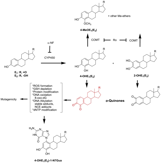
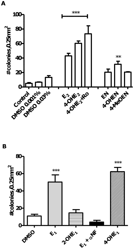
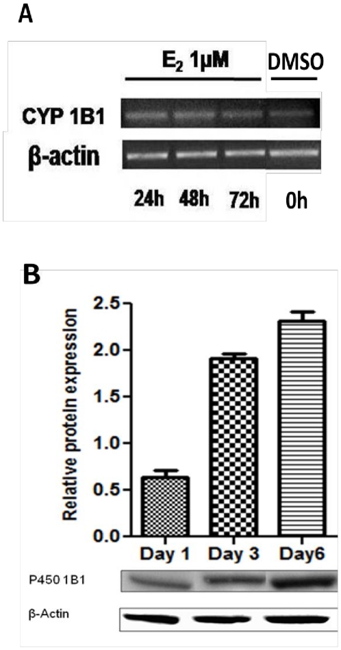
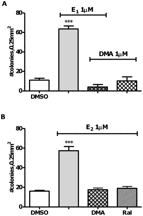
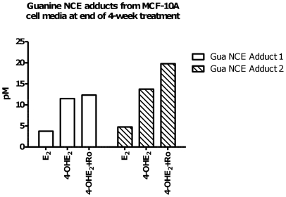
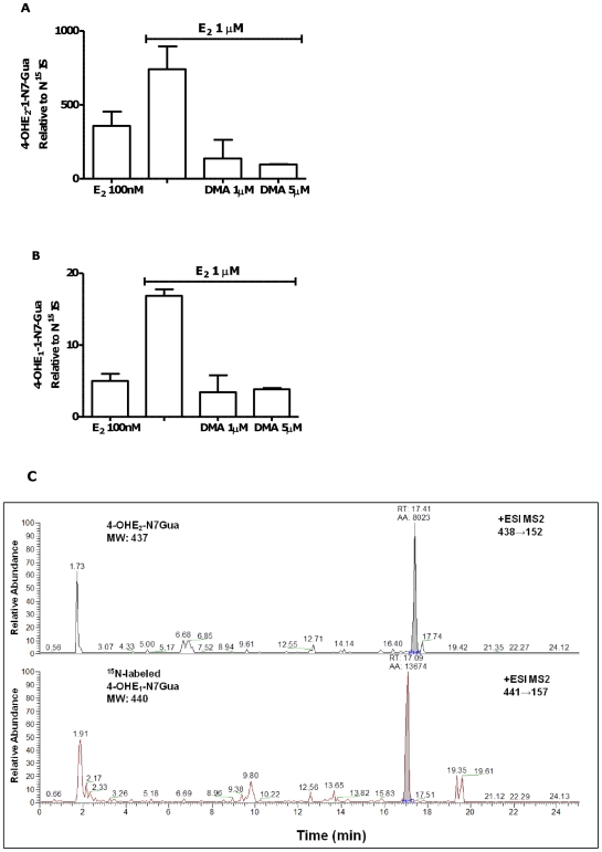
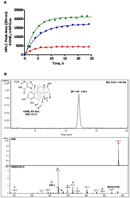
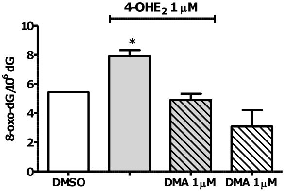
There is association between exposure to estrogens and the development and progression of hormone-dependent gynecological cancers. Chemical carcinogenesis by catechol estrogens derived from oxidative metabolism is thought to contribute to breast cancer, yet exact mechanisms remain elusive. Malignant transformation was studied in MCF-10A human mammary epithelial cells, since estrogens are not proliferative in this cell line. The human and equine estrogen components of estrogen replacement therapy (ERT) and their catechol metabolites were studied, along with the influence of co-administration of selective estrogen receptor modulators (SERMs), raloxifene and desmethyl-arzoxifene (DMA), and histone deacetylase inhibitors. Transformation was induced by human estrogens, and selectively by the 4-OH catechol metabolite, and to a lesser extent by an equine estrogen metabolite. The observed estrogen-induced upregulation of CYP450 1B1 in estrogen receptor negative MCF-10A cells, was compatible with a causal role for 4-OH catechol estrogens, as was attenuated transformation by CYP450 inhibitors. Estrogen-induced malignant transformation was blocked by SERMs correlating with a reduction in formation of nucleobase catechol estrogen (NCE) adducts and formation of 8-oxo-dG. NCE adducts can be formed consequent to DNA abasic site formation, but NCE adducts were also observed on incubation of estrogen quinones with free nucleotides. These results suggest that NCE adducts may be a biomarker for cellular electrophilic stress, which together with 8-oxo-dG as a biomarker of oxidative stress correlate with malignant transformation induced by estrogen oxidative metabolites. The observed attenuation of transformation by SERMs correlated with these biomarkers and may also be of clinical significance in breast cancer chemoprevention.


Conflict of interest statement

Competing Interests: The authors have declared that no competing interests exist.

Figures

Figure 1
Figure 1. Estrogens (E1 and E2) undergo oxidative metabolism to C2 and C4 catechols.
Catechols are further oxidized to form o-quinones. Based on t1/2 and reactivity, the most mutagenic species is 4-OHE1/2 and its further oxidized species. 4-OHE-3,4-o-quinone (shown in red) can redox cycle to form ROS, oxidize DNA (measured as 8-oxo-dG), alkylate DNA (form abasic sites, and stable adducts), and chemically modify other important macromolecules such as dNTP, GSH, and cellular proteins.
Figure 2
Figure 2. Malignant transformation induced by endogenous and equine estrogens: MCF-10A cellular transformation induced by estrogens and metabolites was measured as anchorage-independent growth in soft agar after four weeks.
A. Cells were treated with estrogens or metabolites (1 µM) and COMT inhibitor Ro 41-0960 (Ro, 3 µmol) for 4 weeks before transfer to soft agar. Using one-way ANOVA with Dunnett's post test: *** p<0.001; ** p<0.01 versus DMSO control. B. MCF-10A cellular transformation induced by E1 with or without the CYP450 1B1 inhibitor αNF (3 µM), and a comparison between transformation potency of 2-OHE1 versus 4-OHE1 in MCF-10A cells, 1 µM each. Using one-way ANOVA with Dunnett's post test: *** p<0.001; ** p<0.01 versus DMSO control.
Figure 3
Figure 3. Induction of CYP450 1B1 expression in MCF-10A cells by E2 (1 µM).
Cells were collected at different time points (24 h, 2, 3 and 6 days). Increased CYP450 1B1 mRNA levels A. from E2 treatment measured with real-time PCR, correlate with protein levels B. extracted and analyzed by western blot using anti CYP450 1B1 rabbit polyclonal primary antibody. Representative blots are shown of the 55 kDa immunoreactive band. Intensity of the bands was normalized to β-actin (N = 6) showing mean and s.e.m.
Figure 4
Figure 4. Malignant transformation induced by endogenous estrogens is inhibited by benzothiophene SERMs, but not by HDAC inhibitors.
MCF-10A cellular transformation induced by E1 or E2 in the presence of benzothiophene SERMs (1 µM) was measured as anchorage-independent growth in soft agar after four weeks. A. DMA or B. DMA and the structurally related SERM, raloxifene. Cells were treated for four weeks before transfer to soft agar. Using one-way ANOVA with Dunnett's post test: *** p<0.001 versus DMSO control.
Figure 5
Figure 5. Biomarker study of guanine NCE adducts measured in supernatant of cells treated with E2 and catechol metabolites; guanine adducts correlate with transformation potency.
Guanine NCE adducts of 4-OHE1 were measured in media from MCF-10A cells treated with: E2 1 µM, 4-OHE2 1 µM, or (4-OHE2 1 µM+Ro 3 µM) at week 4. These NCEs adducts were not detectable in vehicle control experiments. N7-Me-guanine (5 nM) was used as internal standard. MRM fragmentation patterns of NCEs (m/z→m/z) corresponded to: Gua NCE Adduct 1 4-OHE1-Gua (436→152); Gua NCE Adduct 2 4-OHE2-Gua adduct (438→152).
Figure 6
Figure 6. Formation of guanine NCE adducts from MCF-10A cells treated with E2 (0.1 or 1 µM) is inhibited by co-treatment with DMA.
Cell culture supernatant was analyzed after 4 days of treatment and 1 week post cell-plating. LC-MS/MS peak area for A. 4-OHE2-1-N7Gua and B. 4-OHE1-1-N7Gua was normalized to equal amounts of internal standard 15N-labeled Gua adduct. C. Representative chromatograms of guanine NCE adducts obtained with the TSQ MS instrument. Guanine adduct 4-OHE2-1-N7Gua from MCF-10A cell media treated with 1 µM E2 after 3 days. 15N-labeled guanine adduct shown in red is the internal standard.
Figure 7
Figure 7. Formation of 4-OHE2-1-N7Gua from reaction of E2-3,4-Q with free Gua nucleotides.
A. Reaction of quinone (0.2 mM) with dGMP (red), dGDP (blue), or dGTP (green) (1.9 mM) buffered at pH 6.8 (phosphate, 10 mM). Data are fit to a pseudo-first order exponential. B. Representative chromatogram of 4-OHE2-1-N7Gua.
Figure 8
Figure 8. 4-OHE2 induced oxidative damage in MCF-10A cells and DMA prevents the formation of 8-oxo-dG.
Cells were treated with 1 µM 4-OHE2 with or without DMA 1 µM for 72 h. 8-Oxo-dG was detected and quantified by LC-MS/MS and dG was measured by HPLC. Data show mean and s.e.m. p<0.05 versus vehicle control and DMA co-treated group by ANOVA with Tukey's post test.

Similar articles

Cited by

References

    1. Russo J, Hasan Lareef M, Balogh G, Guo S, et al. Estrogen and its metabolites are carcinogenic agents in human breast epithelial cells. J Steroid Biochem Mol Biol. 2003;87:1–25. - PubMed
    1. Liehr JG. Is estradiol a genotoxic mutagenic carcinogen? Endocr Rev. 2000;21:40–54. - PubMed
    1. Rossouw JE, Anderson GL, Prentice RL, LaCroix AZ, Kooperberg C, et al. Risks and benefits of estrogen plus progestin in healthy postmenopausal women: principal results From the Women's Health Initiative randomized controlled trial. JAMA. 2002;288:321–333. - PubMed
    1. Beral V. Breast cancer and hormone-replacement therapy in the Million Women Study. Lancet. 2003;362:419–427. - PubMed
    1. Ravdin PM, Cronin KA, Howlader N, Berg CD, Chlebowski RT, et al. The decrease in breast-cancer incidence in 2003 in the United States. N Engl J Med. 2007;356:1670–1674. - PubMed

Publication types

MeSH terms