. 2011;6(11):e28050.
doi: 10.1371/journal.pone.0028050. Epub 2011 Nov 30.

Altered insulin receptor signalling and β-cell cycle dynamics in type 2 diabetes mellitus

Affiliations

Altered insulin receptor signalling and β-cell cycle dynamics in type 2 diabetes mellitus

Franco Folli et al. PLoS One. 2011.

Abstract

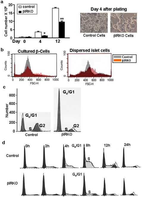
Insulin resistance, reduced β-cell mass, and hyperglucagonemia are consistent features in type 2 diabetes mellitus (T2DM). We used pancreas and islets from humans with T2DM to examine the regulation of insulin signaling and cell-cycle control of islet cells. We observed reduced β-cell mass and increased α-cell mass in the Type 2 diabetic pancreas. Confocal microscopy, real-time PCR and western blotting analyses revealed increased expression of PCNA and down-regulation of p27-Kip1 and altered expression of insulin receptors, insulin receptor substrate-2 and phosphorylated BAD. To investigate the mechanisms underlying these findings, we examined a mouse model of insulin resistance in β-cells--which also exhibits reduced β-cell mass, the β-cell-specific insulin receptor knockout (βIRKO). Freshly isolated islets and β-cell lines derived from βIRKO mice exhibited poor cell-cycle progression, nuclear restriction of FoxO1 and reduced expression of cell-cycle proteins favoring growth arrest. Re-expression of insulin receptors in βIRKO β-cells reversed the defects and promoted cell cycle progression and proliferation implying a role for insulin-signaling in β-cell growth. These data provide evidence that human β- and α-cells can enter the cell-cycle, but proliferation of β-cells in T2DM fails due to G1-to-S phase arrest secondary to defective insulin signaling. Activation of insulin signaling, FoxO1 and proteins in β-cell-cycle progression are attractive therapeutic targets to enhance β-cell regeneration in the treatment of T2DM.


Conflict of interest statement

Competing Interests: The authors have declared that no competing interests exist.

Figures

Figure 1
Figure 1. Reduced β-cell mass, enhanced PCNA+ β- and α-cells and altered expression of insulin signaling and cell adhesion proteins in pancreas from patients with T2DM.
Representative confocal mages of pancreas sections from type 2 diabetic and control pancreas showing (a) quantification of islet cell number with immunostaining for insulin (red), glucagon (blue) and somatostatin (red); PCNA+ cells co-staining with (b) insulin or glucagons (red) and PCNA (green) and (c) CK19 (green), PCNA (red) and insulin (blue); (d) alterations in expression of β-catenin with immunstaining for PCNA (blue), β-catenin (green), insulin (red), light blue in the merged image indicates co-localization between β-catenin and PCNA; (e) alterations in E-cadherin with immunostaining for PCNA (blue), E-cadherin (green) and insulin (red) and the merged image in yellow; Immunostaining of pancreas sections for (f) insulin receptor α-subunit (green), insulin (red) and glucagon (blue); (g) BAD (green) and insulin (red) and merged image in yellow; (h) phospho-BAD (green), insulin (red) and merged image in yellow, and (i) the cell-cycle inhibitor p27-kip1 (green), insulin (red) and glucagon (blue), (n = 7). Yellow in merged images indicates co-localization between insulin staining and insulin signalling proteins. Immunostaining was performed as described in Methods. Bars = 50 µm. *, p<0.05 controls vs T2D.
Figure 2
Figure 2. Reduced transcript levels of insulin signaling and cell-cycle proteins and protein levels of insulin signaling components in islets isolated from patients with T2DM.
(a) Expression of insulin receptor isoforms A and B; (b) transcript levels of insulin signaling proteins, and (c) transcript levels of cell-cycle proteins. Data are expressed after normalization for TATA binding protein (TBP) (n = 5–7); *, p<0.05; ***, p<0.001; controls vs T2DM. (d) protein expression of total insulin receptors, (e), total insulin receptor substrate-1, (f) total insulin receptor substrate-2, (g), tyrosine-612 IRS-1, (h) tyrosine-612 IRS-2, (i) or total phosphatidyl inositol 3-kinase. *,p<0.05; **,p<0.001 Control vs T2D. Data in 2d–i are immunoprecipitates. (j) β-actin was used to normalize the data in Figures 2d–i.
Figure 3
Figure 3. Insulin receptor-deficient β-cells exhibit slow growth and block in G1 to S-phase transition.
(a) Defects in growth of βIRKO cells is evident from total number of cells 4 and 12 days after the same number of cells are plated at day 0; the panel on the right shows poor spread and clumping of βIRKO cells (b) β-cell size in control and βIRKO cell lines and islets determined by FACS analyses with forward scatter; (c) significantly lower G1 and G2 in βIRKO cells, and (d) block in G1 to S-phase transition in βIRKO cells after synchronization with hydroxyurea for 18h. Analyses were performed by FACS. In each case, representative data from 3 clones each derived from 3 RIP-Cre controls and 3 βIRKO mice are shown.
Figure 4
Figure 4. Insulin receptor-deficient β-cells show reduced expression of cyclins and cyclin dependent kinases and nuclear restriction of FoxO1.
(a) Reduced expression of cdk2 and cdk4; (b) reduced expression of cyclin D2, cyclin D3 and cyclin E; (c) increased expression and nuclear restriction of FoxO1; (d) re-expression of the insulin receptor in βIRKO β-cells (βIRKO+hIRB) restores the expression of CDK2, CDK4 and cyclin E. Actin is used as a loading control; (e) reduced p-IRS2 and p-S473-Akt in βIRKO and restoration of total IRS2, pIRS2 and p-S473-Akt in insulin receptor re-expressing cells (βIRKO+hIRB); (f) Re-expression of insulin receptors in βIRKO cells restores FoxO1 to nucleus; (g) proliferation analyzed by the deuterated water technique . Representative data from at least 3 independent Western blotting experiments from at least 2 independent clones are shown. The plots below the blots are shown in arbitrary units. The loading control for experiments in Fig. 4a–c is presented under the blot for cyclin D3 in Fig. 4b. *, p<0.05 βIRKO vs Control or βIRKO+hIRB.
Figure 5
Figure 5. Schematic linking the growth factor pathway to defects in cell cycle progression and apoptosis in diabetic β-cells.

References

    1. Accili D. Lilly lecture 2003: the struggle for mastery in insulin action: from triumvirate to republic. Diabetes. 2004;53:1633–1642. - PubMed
    1. DeFronzo RA. Pathogenesis of type 2 diabetes mellitus. Med Clin North Am. 2004;88:787–835. - PubMed
    1. Saltiel AR, Kahn CR. Insulin signalling and the regulation of glucose and lipid metabolism. Nature. 2001;414:799–806. - PubMed
    1. Kawamori D, Kurpad AJ, Hu J, Liew CW, Shih JL, et al. Insulin signaling in alpha cells modulates glucagon secretion in vivo. Cell Metab. 2009;9:350–361. - PMC - PubMed
    1. Ling C, Groop L. Epigenetics: a molecular link between environmental factors and type 2 diabetes. Diabetes. 2009;58:2718–2725. - PMC - PubMed

Publication types

MeSH terms