. 2011;6(11):e28131.
doi: 10.1371/journal.pone.0028131. Epub 2011 Nov 30.

Type I interferon reaction to viral infection in interferon-competent, immortalized cell lines from the African fruit bat Eidolon helvum

Affiliations

Type I interferon reaction to viral infection in interferon-competent, immortalized cell lines from the African fruit bat Eidolon helvum

Susanne E Biesold et al. PLoS One. 2011.

Abstract

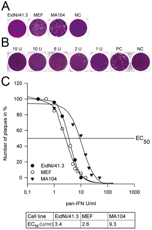
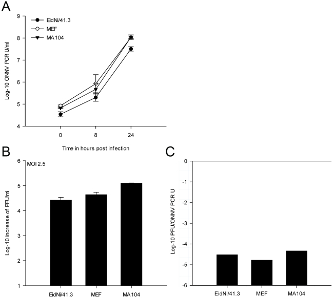
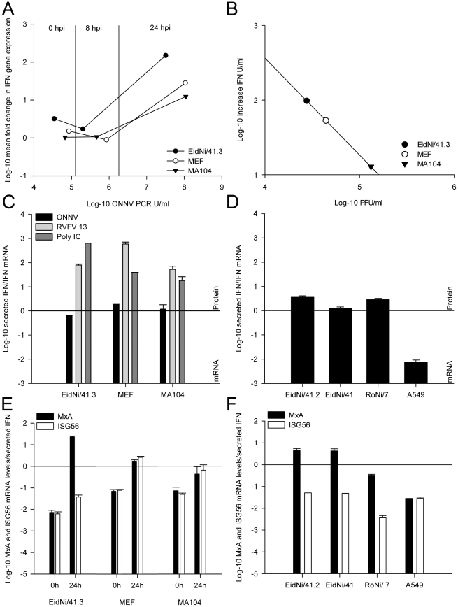
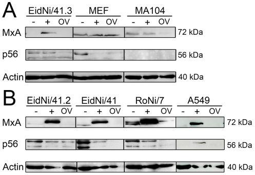
Bats harbor several highly pathogenic zoonotic viruses including Rabies, Marburg, and henipaviruses, without overt clinical symptoms in the animals. It has been suspected that bats might have evolved particularly effective mechanisms to suppress viral replication. Here, we investigated interferon (IFN) response, -induction, -secretion and -signaling in epithelial-like cells of the relevant and abundant African fruit bat species, Eidolon helvum (E. helvum). Immortalized cell lines were generated; their potential to induce and react on IFN was confirmed, and biological assays were adapted to application in bat cell cultures, enabling comparison of landmark IFN properties with that of common mammalian cell lines. E. helvum cells were fully capable of reacting to viral and artificial IFN stimuli. E. helvum cells showed highest IFN mRNA induction, highly productive IFN protein secretion, and evidence of efficient IFN stimulated gene induction. In an Alphavirus infection model, O'nyong-nyong virus exhibited strong IFN induction but evaded the IFN response by translational rather than transcriptional shutoff, similar to other Alphavirus infections. These novel IFN-competent cell lines will allow comparative research on zoonotic, bat-borne viruses in order to model mechanisms of viral maintenance and emergence in bat reservoirs.


Conflict of interest statement

Competing Interests: The authors have declared that no competing interests exist.

Figures

Figure 1
Figure 1. Generating immortalized bat cell cultures and measuring interferon (IFN)-β mRNA induction.
(A) Primary bat cell cultures were generated from an embryonic kidney of E. helvum. Cell cultures were immortalized by lentiviral transduction of a simian virus 40 large T antigen. Three clonal cell lines (EidNi/41.1, 41.2 and 41.3) were prepared from a mixed culture (EidNi/41) by end point dilution plating. Bars indicate 20 µm. (B) Phylogenetic relationships of different selected bat species for the generation of target gene sequences (adapted from [3]). For the IFN-β gene four species were selected. (C) Alignment of bat IFN-β genes (E. helvum, M. daubentonii, R. cf. landeri, R. aegyptiacus) and positions of primers and probe for a pan-bat real-time RT-PCR. (D) Replication of a Renilla luciferase expressing Rift valley fever virus clone 13 (RVFV 13) in various cell lines (EidNi/41.3, MEF (mouse embryonic fibroblasts), MA104 (African green monkey kidney) and as reference Vero cells at different MOIs. Cells were infected at different ten-fold diluted MOIs (7.5 until 0.000075) and lysed 24 hpi with Renilla lysis buffer (Promega). Replication was detected by Renilla luciferase read-out. Experiments were performed in duplicates. Highest replication was found in Vero cells followed by EidNi/41.3, MEF, MA104 (between10 to 100-fold less compared to Vero cells). (E) IFN-β mRNA transcription was induced by either RVFV 13 infection (MOI 1) or poly IC transfection (5 µg per 6-well). IFN-β and TATA-box binding protein (housekeeping gene) mRNA was quantified by species-specific real-time RT-PCR assays. The fold induction was calculated with the 2−ΔΔCt method.
Figure 2
Figure 2. Interferon quantification and calibration by vesicular stomatitis virus (VSV) bioassay.
(A) VSV plaque morphology was analyzed on the bat cell line EidNi/41.3, a rodent cell line (MEF) and a primate cell line (MA104). (B) For the VSV bioassay EidNi/41.3 cells were pre-incubated with different amounts (units per ml; U/ml) of pan-species IFN (pan-IFN). 24 h after treatment the cells were infected with VSV at an MOI of 0.025 for 1 h. After 2 days cells were fixed, stained and plaques were counted to estimate the correlation between the amounts of pan-IFN and plaques. (C) For each cell line a standard curve using different amount of pan-IFN was done and EC50 values were calculated. Shown are mean values of quadruplicates. Standard deviations are not shown for clarity.
Figure 3
Figure 3. Bat cells produce high levels of secreted IFN.
Cells were infected with RVFV 13 or transfected with poly IC as described before. With the help of the VSV bioassay secreted IFN was measured. Each cell line was incubated for 24 h with IFN-containing supernatants (β-propiolactone inactivated) and with pan-IFN standards diluted in medium from untreated control cells. IFN concentrations were normalized with the help of EC50 values as described in the Methods section.
Figure 4
Figure 4. O'nyong-nyong virus (ONNV) replication in different mammalian cells using a high MOI.
(A) For a synchronized infection cells were inoculated with ONNV at an MOI 2.5 and supernatants were harvested at 0, 8 and 24 h post infection (hpi). After viral RNA isolation (triplicates) the concentration was measured by ONNV specific real-time RT-PCR assay. ONNV PCR units (U) per ml were determined. The dilution end-point was defined as one PCR unit. Virus replication could be detected in all cell lines. The increase of genome equivalents per ml were approximately 1000-fold after 24 hpi. (B) Titration of supernatants showed an increase of PFU per ml (titer of inoculum was subtracted) after 24 hpi of 1000 to 10000-fold. (C) The ratio of log-10 increase PFU/ml to ONNV PCR units were comparable in all cell lines indicating an efficient particle formation.
Figure 5
Figure 5. IFN-β mRNA induction but IFN protein decrease in all mammalian cells upon ONNV infection.
(A) IFN-β mRNA induction was measured by species-specific real-time RT-PCR at time points 0, 8 and 24 hpi and correlated to the amount of relative ONNV genome equivalents in PCR units per ml. ONNV replication led to an induction of IFN-β mRNA 24 hpi. (B) At 24 hpi the increase of secreted IFN was correlated to ONNV plaque forming units indicating that higher virus titres led to a decreased amount of IFN in the supernatants. (C) Comparison of secreted IFN protein to IFN-β mRNA (24 hpi) after ONNV, RVFV 13 infection and poly IC transfection. ONNV replication was related to IFN protein reduction. For absolute values refer to Figure S1. (D) Confirmation for IFN protein reduction by testing different EidNi bat cell cultures. EidNi/41.2 (subclone), EidNi/41 (mixed cell culture), RoNi/7 (mixed cell culture from R. aegyptiacus kidneys) and human lung adenocarcinoma epithelial cell line (A549) showed the same phenotype. (E) Comparison of mRNA fold-induction of IFN stimulated genes (MxA and ISG56) to secreted IFN protein at time points 0 hpi and 24 hpi. Expression of ISGs was not affected by IFN protein downregulation indicating that there was no general transcriptional shutoff. (F) Confirmation by testing different cell clones and cell cultures 24 hpi indicating that MxA mRNA is upregulated in bat cell cultures upon ONNV infection.
Figure 6
Figure 6. ONNV infection ablates the expression of IFN stimulated genes.
(A) Cells were either left untreated (−) or infected with RVFV 13 (+) or ONNV (OV). After 24 h proteins were extracted from cells. Same amount of proteins were subjected to SDS-PAGE followed by a Western blot analysis. Mouse-anti-Mx1/2/3 (MxA), goat-anti-IFIT1/ISG56 (p56) and mouse-anti-actin immunoglobulins were applied at dilutions 1∶1000 followed by a peroxidase labeled goat-anti-mouse or rabbit-anti-goat secondary antibody (1∶20000). In all cell lines infection with ONNV did not induce the expression of MxA and p56 and was less or comparable to untreated cells. (B) To exclude cell clone specific effects additional EidNi and RoNi bat cell cultures were included (EidNi/41.2; EidNi/41 and RoNi/7) as well as a human A549 cell line.
Figure 7
Figure 7. Efficient ONNV replication upon infection at a low MOI.
(A) In order to analyze if insufficient viral replication led to a delayed or ablated IFN production in cells a growth kinetic at low MOI was performed (MOI 0.0025). Cells were inoculated with ONNV and supernatants were analyzed at 0, 8 and 24 hpi by real-time RT-PCR. Virus replication could be detected in all cell lines. (B) Supernatants were titrated after 24 hpi confirming the PCR results. (C) PFU to ONNV PCR unit (U) ratio after infection of cells with ONNV at an MOI of 0.0025. In MA104 and MEF cells the infection at low MOI resulted in a higher PFU to genome equivalent ratio compared with infection at high MOI.

Similar articles

Cited by

References

    1. Simmons NB. Evolution. An Eocene big bang for bats. Science. 2005;307:527–528. - PubMed
    1. Teeling EC, Springer MS, Madsen O, Bates P, O'Brien S J, et al. A molecular phylogeny for bats illuminates biogeography and the fossil record. Science. 2005;307:580–584. - PubMed
    1. Jones G, Teeling EC. The evolution of echolocation in bats. Trends in Ecology & Evolution. 2006;21:149–156. - PubMed
    1. Calisher CH, Childs JE, Field HE, Holmes KV, Schountz T. Bats: important reservoir hosts of emerging viruses. Clin Microbiol Rev. 2006;19:531–545. - PMC - PubMed
    1. Swanepoel R, Leman PA, Burt FJ, Zachariades NA, Braack LE, et al. Experimental inoculation of plants and animals with Ebola virus. Emerg Infect Dis. 1996;2:321–325. - PMC - PubMed

Publication types

MeSH terms