. 2011;6(11):e28147.
doi: 10.1371/journal.pone.0028147. Epub 2011 Nov 29.

Diverse roles of Eph/ephrin signaling in the mouse lens

Affiliations

Diverse roles of Eph/ephrin signaling in the mouse lens

Catherine Cheng et al. PLoS One. 2011.

Abstract

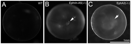
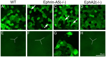
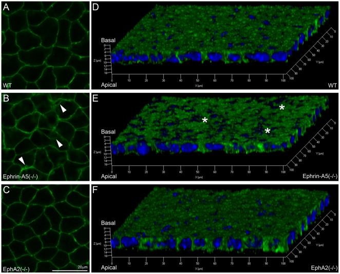
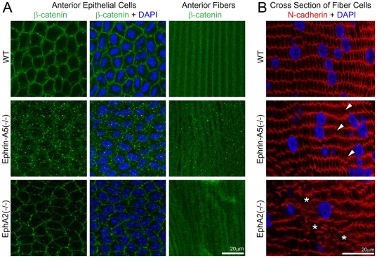
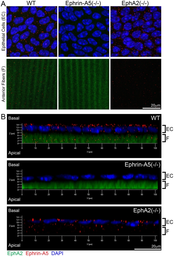
Recent genetic studies show that the Eph/ephrin bidirectional signaling pathway is associated with both congenital and age-related cataracts in mice and humans. We have investigated the molecular mechanisms of cataractogenesis and the roles of ephrin-A5 and EphA2 in the lens. Ephrin-A5 knockout ⁻/⁻ mice often display anterior polar cataracts while EphA2⁻/⁻ lenses show very mild cortical or nuclear cataracts at weaning age. The anterior polar cataract of ephrin-A5⁻/⁻ lenses is correlated with multilayers of aberrant cells that express alpha smooth muscle actin, a marker for mesenchymal cells. Only select fiber cells are altered in ephrin-A5⁻/⁻ lenses. Moreover, the disruption of membrane-associated β-catenin and E-cadherin junctions is observed in ephrin-A5⁻/⁻ lens central epithelial cells. In contrast, EphA2⁻/⁻ lenses display normal monolayer epithelium while disorganization is apparent in all lens fiber cells. Immunostaining of ephrin-A5 proteins, highly expressed in lens epithelial cells, were not colocalized with EphA2 proteins, mainly expressed in lens fiber cells. Besides the previously reported function of ephrin-A5 in lens fiber cells, this work suggests that ephrin-A5 regulates β-catenin signaling and E-cadherin to prevent lens anterior epithelial cells from undergoing the epithelial-to-mesenchymal transition while EphA2 is essential for controlling the organization of lens fiber cells through an unknown mechanism. Ephrin-A5 and EphA2 likely interacting with other members of Eph/ephrin family to play diverse functions in lens epithelial cells and/or fiber cells.


Conflict of interest statement

Competing Interests: The authors have declared that no competing interests exist.

Figures

Figure 1
Figure 1. Different types of cataracts occur in ephrin-A5(-/-) and EphA2(-/-) mice.
Photos of enucleated fresh lenses from P21 wild-type (WT) (A), ephrin-A5(-/-) (B) and EphA2(-/-) (C) mice. A representative ephrin-A5(-/-) lens photo shows an obvious cataract at the anterior pole (arrowhead in B). A representative EphA2(-/-) lens displays mild nuclear opacity (arrowheads in C). Scale bar, 1mm.
Figure 2
Figure 2. Confocal images of the anterior region of GFP+ wild-type (WT), ephrin-A5(-/-) and EphA2 lenses.
The WT lens shows typical mosaic GFP expression pattern in the central epithelium (A) and the Y-shape suture (Y-line) of underlying fiber cells (E). An ephrin-A5(-/-) lens displays morphological changes in a few epithelial cells (arrows in B), and the other ephrin-A5(-/-) lens shows a cluster of aberrant cells underneath the central epithelial cells (arrows in C). In addition, mislocalized aberrant cells are apparent in underlying fiber cell layers without the normal Y-shaped suture (G). The EphA2 (-/-) lens has normal central epithelial cells (D) and anterior Y-shaped suture (H). Scale bar, 50 µm.
Figure 3
Figure 3. Characterization of lens anterior polar regions in GFP+ WT and ephrin-A5(-/-) lenses.
Three-dimensional reconstruction of a P21 GFP+ ephrin-A5(-/-) lens reveals a large cluster of aberrant cells underneath anterior epithelial cells (A). Scale bar, 50 µm. Immunostaining of α-SMA (red) in anterior epithelial cells from lens capsule flat mounts of P21 WT (B) and ephrin-A5(-/-) lenses (C). Only ephrin-A5 (-/-) anterior epithelial cells that are abnormally clustered in the fiber cell layer are obviously positive for α-SMA. Scale bar, 20 µm. Three-dimensional reconstruction of the anterior polar region of P14 GFP+ WT (D) and ephrin-A5(-/-) lenses (F). Scale bar, 50 µm. Enlarged views of anterior epithelial cells in D and F are the boxed areas in panels E and G. Scale bar, 20 µm.
Figure 4
Figure 4. E-cadherin distribution in WT, ephrin-A5(-/-) and EphA2(-/-) lens capsule flat mounts.
Fluorescent images reveal normal staining signals of E-cadherin in WT and EphA2(-/-) anterior epithelial cells (A and C) but alterations in the ephrin-A5(-/-) anterior epithelium (B, indicated by arrowheads). Three-dimensional reconstructions of z-stack images labeled for E-cadherin (green) and DAPI (blue, nuclei) of lens epithelial cells from P21 WT, ephrin-A5(-/-) and EphA2(-/-) lens capsule flat mounts (D, E and F). There is a notable disruption of E-cadherin staining in ephrin-A5(-/-) anterior epithelial cells as compared to those in WT and EphA2(-/-) cells. Scale bar, 20 µm.
Figure 5
Figure 5. Beta-catenin distribution in anterior epithelial cells and underlying lens fibers and N-cadeherin localization in lens frozen sections.
Immunostaining of lens capsule flat mounts shows cell membrane distribution of β-catenin proteins in both anterior epithelial cells and fiber cells of a P21 WT lens (A). Membrane association of β-catenin remains unchanged in EphA2(-/-) anterior lens epithelial cells, but β-catenin proteins form substantial small aggregates in both anterior epithelial cells and fiber cells of ephrin-A5(-/-) lenses (A). Scale bar, 20 µm. N-cadherin is localized at the cell boundaries of hexagonal shaped lens fibers in the WT lens section (B). In the ephrin-A5(-/-) lens section, the majority of fiber cells show normal localization of N-cadherin proteins except select abnormal fiber cells (B, arrowheads). In the EphA2(-/-) lens section, the distribution of N-cadherin proteins is severely altered with some areas lacking N-cadherin staining (B, asterisks). Scale bar, 20 µm.
Figure 6
Figure 6. Localization of ephrin-A5 and EphA2 in the lens.
Double immunolabeling of ephrin-A5 (red) and EphA2 (green) with DAPI staining (blue, nuclei) of anterior lens epithelial and fiber cells from lens capsule flat mounts of P21 wild-type (WT) and ephrin-A5(-/-), EphA2(-/-) mice (A). Side views of z-stack reconstructions of anterior epithelial cells with underlying fiber cells of P21 WT, ephrin-A5(-/-), EphA2(-/-) lens capsule flat mounts reveal that ephrin-A5 proteins (red) show punctate signals mostly at the lateral and apical sides of lens epithelial cells (EC), and EphA2 proteins (green) show diffused signals in fiber cells (F) (B). Scale bar, 20 µm.

References

    1. Yamamoto N, Majima K, Marunouchi T. A study of the proliferating activity in lens epithelium and the identification of tissue-type stem cells. Med Mol Morphol. 2008;41:83–91. - PubMed
    1. Piatigorsky J. Lens differentiation in vertebrates. A review of cellular and molecular features. Differentiation. 1981;19:134–153. - PubMed
    1. Zampighi GA, Eskandari S, Kreman M. Epithelial organization of the mammalian lens. Exp Eye Res. 2000;71:415–435. - PubMed
    1. Kuszak JR, Novak LA, Brown HG. An ultrastructural analysis of the epithelial-fiber interface (EFI) in primate lenses. Exp Eye Res. 1995;61:579–597. - PubMed
    1. Bassnett S, Winzenburger PA. Morphometric analysis of fibre cell growth in the developing chicken lens. Exp Eye Res. 2003;76:291–302. - PubMed

Publication types

MeSH terms