

ERG deregulation induces PIM1 over-expression and aneuploidy in prostate epithelial cells
- PMID: 22140532
- PMCID: PMC3227636
- DOI: 10.1371/journal.pone.0028162
ERG deregulation induces PIM1 over-expression and aneuploidy in prostate epithelial cells
Abstract
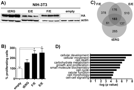
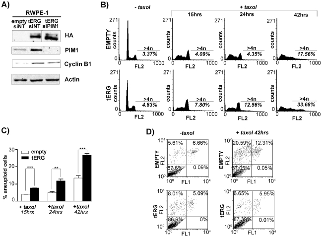
The ERG gene belongs to the ETS family of transcription factors and has been found to be involved in atypical chromosomal rearrangements in several cancers. To gain insight into the oncogenic activity of ERG, we compared the gene expression profile of NIH-3T3 cells stably expressing the coding regions of the three main ERG oncogenic fusions: TMPRSS2/ERG (tERG), EWS/ERG and FUS/ERG. We found that all three ERG fusions significantly up-regulate PIM1 expression in the NIH-3T3 cell line. PIM1 is a serine/threonine kinase frequently over-expressed in cancers of haematological and epithelial origin. We show here that tERG expression induces PIM1 in the non-malignant prostate cell line RWPE-1, strengthening the relation between tERG and PIM1 up-regulation in the initial stages of prostate carcinogenesis. Silencing of tERG reversed PIM1 induction. A significant association between ERG and PIM1 expression in clinical prostate carcinoma specimens was found, suggesting that such a mechanism may be relevant in vivo. Chromatin Immunoprecipitation experiments showed that tERG directly binds to PIM1 promoter in the RWPE-1 prostate cell line, suggesting that tERG could be a direct regulator of PIM1 expression. The up-regulation of PIM1 induced by tERG over-expression significantly modified Cyclin B1 levels and increased the percentage of aneuploid cells in the RWPE-1 cell line after taxane-based treatment. Here we provide the first evidence for an ERG-mediated PIM1 up-regulation in prostate cells in vitro and in vivo, suggesting a direct effect of ERG transcriptional activity in the alteration of genetic stability.
Conflict of interest statement
Figures


Similar articles
-
Pim1 promotes human prostate cancer cell tumorigenicity and c-MYC transcriptional activity.BMC Cancer. 2010 Jun 1;10:248. doi: 10.1186/1471-2407-10-248. BMC Cancer. 2010. PMID: 20515470 Free PMC article.
-
Binding of TMPRSS2-ERG to BAF Chromatin Remodeling Complexes Mediates Prostate Oncogenesis.Mol Cell. 2018 Aug 16;71(4):554-566.e7. doi: 10.1016/j.molcel.2018.06.040. Epub 2018 Aug 2. Mol Cell. 2018. PMID: 30078722 Free PMC article.
-
Abnormal expression of the ERG transcription factor in prostate cancer cells activates osteopontin.Mol Cancer Res. 2011 Jul;9(7):914-24. doi: 10.1158/1541-7786.MCR-10-0537. Epub 2011 Jun 13. Mol Cancer Res. 2011. PMID: 21669963
-
[The progress of TMPRSS2-ETS gene fusions and their mechanism in prostate cancer].Yi Chuan. 2011 Feb;33(2):117-22. doi: 10.3724/sp.j.1005.2011.00117. Yi Chuan. 2011. PMID: 21377967 Review. Chinese.
-
The oncogene ERG: a key factor in prostate cancer.Oncogene. 2016 Jan 28;35(4):403-14. doi: 10.1038/onc.2015.109. Epub 2015 Apr 27. Oncogene. 2016. PMID: 25915839 Review.
Cited by
-
The Novel PIM1 Inhibitor NMS-P645 Reverses PIM1-Dependent Effects on TMPRSS2/ERG Positive Prostate Cancer Cells And Shows Anti-Proliferative Activity in Combination with PI3K Inhibition.J Cancer. 2017 Jan 1;8(1):140-145. doi: 10.7150/jca.15838. eCollection 2017. J Cancer. 2017. PMID: 28123608 Free PMC article.
-
Emergence of ETS transcription factors as diagnostic tools and therapeutic targets in prostate cancer.Am J Transl Res. 2013 Apr 19;5(3):254-68. Print 2013. Am J Transl Res. 2013. PMID: 23634237 Free PMC article.
-
Androgen receptor and gene network: Micromechanics reassemble the signaling machinery of TMPRSS2-ERG positive prostate cancer cells.Cancer Cell Int. 2014 Apr 17;14:34. doi: 10.1186/1475-2867-14-34. eCollection 2014. Cancer Cell Int. 2014. PMID: 24739220 Free PMC article. Review.
-
Expression and ERG regulation of PIM kinases in prostate cancer.Cancer Med. 2021 May;10(10):3427-3436. doi: 10.1002/cam4.3893. Epub 2021 May 1. Cancer Med. 2021. PMID: 33932111 Free PMC article.
-
Covid-19 pathogenesis in prostatic cancer and TMPRSS2-ERG regulatory genetic pathway.Infect Genet Evol. 2021 Mar;88:104669. doi: 10.1016/j.meegid.2020.104669. Epub 2020 Dec 7. Infect Genet Evol. 2021. PMID: 33301988 Free PMC article. Review.
References
-
- Marcucci G, Baldus CD, Ruppert AS, Radmacher MD, Mrozek K, et al. Overexpression of the ETS-related gene, ERG, predicts a worse outcome in acute myeloid leukemia with normal karyotype: a Cancer and Leukemia Group B study. J Clin Oncol. 2005;23:9234–9242. - PubMed
-
- Baldus CD, Burmeister T, Martus P, Schwartz S, Gokbuget N, et al. High expression of the ETS transcription factor ERG predicts adverse outcome in acute T-lymphoblastic leukemia in adults. J Clin Oncol. 2006;24:4714–4720. - PubMed
-
- Delattre O, Zucman J, Plougastel B, Desmaze C, Melot T, et al. Gene fusion with an ETS DNA-binding domain caused by chromosome translocation in human tumours. Nature. 1992;359:162–165. - PubMed
-
- Sorensen PH, Lessnick SL, Lopez-Terrada D, Liu XF, Triche TJ, et al. A second Ewing's sarcoma translocation, t(21;22), fuses the EWS gene to another ETS-family transcription factor, ERG. Nat Genet. 1994;6:146–151. - PubMed
Publication types
MeSH terms
Substances
LinkOut - more resources
Full Text Sources
Molecular Biology Databases
