. 2011 Nov;5(11):e1410.
doi: 10.1371/journal.pntd.0001410. Epub 2011 Nov 29.

Inhibition of dengue virus entry and multiplication into monocytes using RNA interference

Affiliations

Inhibition of dengue virus entry and multiplication into monocytes using RNA interference

Mohammed Abdelfatah Alhoot et al. PLoS Negl Trop Dis. 2011 Nov.

Abstract

Background: Dengue infection ranks as one of the most significant viral diseases of the globe. Currently, there is no specific vaccine or antiviral therapy for prevention or treatment. Monocytes/macrophages are the principal target cells for dengue virus and are responsible for disseminating the virus after its transmission. Dengue virus enters target cells via receptor-mediated endocytosis after the viral envelope protein E attaches to the cell surface receptor. This study aimed to investigate the effect of silencing the CD-14 associated molecule and clathrin-mediated endocytosis using siRNA on dengue virus entry into monocytes.

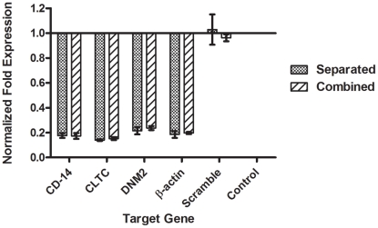
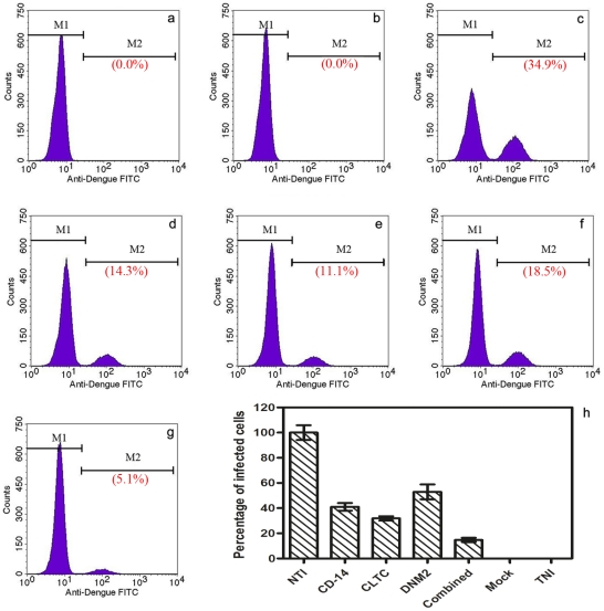
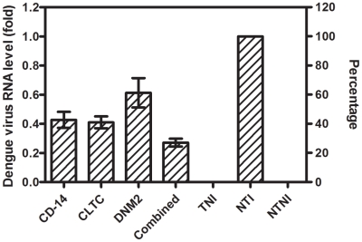
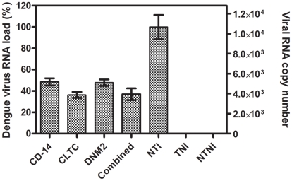
Methodology/principal findings: Gene expression analysis showed a significant down-regulation of the target genes (82.7%, 84.9 and 76.3% for CD-14 associated molecule, CLTC and DNM2 respectively) in transfected monocytes. The effect of silencing of target genes on dengue virus entry into monocytes was investigated by infecting silenced and non-silenced monocytes with DENV-2. Results showed a significant reduction of infected cells (85.2%), intracellular viral RNA load (73.0%), and extracellular viral RNA load (63.0%) in silenced monocytes as compared to non-silenced monocytes.

Conclusions/significance: Silencing the cell surface receptor and clathrin mediated endocytosis using RNA interference resulted in inhibition of the dengue virus entry and subsequently multiplication of the virus in the monocytes. This might serve as a novel promising therapeutic target to attenuate dengue infection and thus reduce transmission as well as progression to severe dengue hemorrhagic fever.


Conflict of interest statement

The authors have declared that no competing interests exist.

Figures

Figure 1
Figure 1. Silencing efficiency of target genes.
siRNA pools were proven to be specific and potent in silencing target genes. Each gene of the target genes was targeted with a pool of siRNAs consisted of three different siRNAs. Results showed an efficient gene knockdown when compared with non-transfected control and normalized to reference genes (One-way ANOVA with Dunnett's post-test, P<0.0001, Error bars are ± SD). The effect of a combined transfection of the siRNA pools in monocytes is consistent, and there are no significant differences between separated transfection, (82.4%±1.9), (86.0%±0.6), and (78.5%±3.0), and combined transfection (82.7%±2.4), (84.9%±1.2), and (76.3%±1.7), on expression of CD-14 associated molecule, CLTC, and DNM2, respectively (Two-way ANOVA with Bonferroni post-test, P>0.05, Error bars are ± SD). Scrambled siRNA had no inhibitory effect on any gene expression and showed a similar expression to the non-transfected control.
Figure 2
Figure 2. Transfection cytotoxicity.
Monocytes were exposed to siRNA at a concentration of 25, 50, and 50 nM for CD-14 associate molecule, CLTC, and DNM2, respectively. Cytotoxicity was determined by measuring LDH level in the culture supernatant and by counting trypan blue stained viable cells. Results showed siRNAs are not cytotoxic (>90% live cells) for all pools of siRNA. Combined transfection of different siRNAs also found not cytotoxic to monocytes (90.5% live cells). No statistical significance was observed by One-way ANOVA analysis (P>0.05).
Figure 3
Figure 3. Quantification of infected cells by flow cytometry.
At 72 h post-transfection, monocytes were infected by DENV-2 at MOI of 2. Marked reduction in percentage of infected cells was observed. This figure shows the percentage of DENV infected cells at different conditions. (a) Transfected non-infected monocytes (0.0%) represents the negative control. (b) Transfected mock-infected monocytes as a staining control (0.0%). (c) Non-transfected infected monocytes (34.9%) as a positive control. (d) CD-14 associated molecule silenced infected monocytes (14.3%). (e) CLTC silenced infected monocytes (11.1%). (f) DNM2 silenced infected monocytes (18.5%). (g) CD-14 associated molecule, CLTC, and DNM2 combined silenced infected monocytes (5.1%). (h) Summarized the results of the flow cytometry experiments. Data is expressed as a percentage of infected cells compared with non-transfected infected monocytes (NTI) which was defined as 100%. The percentages of the infected cells are 41.0%, 36.2%, 52.9% and 14.8% for CD-14 associated molecule, CLTC, DNM2, and combined silenced monocytes, respectively (One-way ANOVA with Dunnett's post-test, P<0.0001, Error bars are ± SD).
Figure 4
Figure 4. Intracellular dengue virus RNA load.
Viral RNA levels were quantified by RT-QPCR and normalized to reference gene (RPL22). Data is expressed as relative fold expression to non-transfected infected monocytes control, which defined as 1.0 fold. Results show (0.57 fold±0.10), (0.59 fold±0.07), (0.39 fold±0.18), and (0.73 fold±0.05) reduction in viral RNA load in CD-14 associated molecule, CLTC, DNM2, and combined silenced monocytes, respectively (One-way ANOVA with Dunnett's post-test, P<0.0001, Error bars are ± SD). (TNI, Transfected Non-Infected; NTI, Non-Transfected Infected; NTNI, Non-Transfected Non-Infected).
Figure 5
Figure 5. Extracellular dengue virus RNA load.
DENV RNA in the culture supernatant of transfected and non-transfected monocytes was quantified by RT-qPCR. Result shows marked reduction in viral RNA (51.4%, 63.7%, 52.2%, and 63.0%) for CD-14 associated molecule, CLTC, DNM2, and combined silenced monocytes, respectively, when compared with non-transfected monocytes, which defined as 100% (viral RNA copy number is 1.06×10⁴/µl). This result is statistically significant (One-way ANOVA with Dunnett's post-test, P<0.0001, Error bars are ± SD). (TNI, Transfected Non-Infected; NTI, Non-Transfected Infected; NTNI, Non-Transfected Non-Infected).

Similar articles

Cited by

References

    1. Smart K, Safitri I. Evidence behind the WHO guidelines: hospital care for children: what treatments are effective for the management of shock in severe dengue? J Trop Pediatr. 2009;55:145–148. - PubMed
    1. Gubler DJ. Dengue and dengue hemorrhagic fever. Clin Microbiol Rev. 1998;11:480–496. - PMC - PubMed
    1. Kurane I, Janus J, Ennis FA. Dengue virus infection of human skin fibroblasts in vitro production of IFN-beta, IL-6 and GM-CSF. Arch Virol. 1992;124:21–30. - PubMed
    1. Hase T, Summers PL, Eckels KH. Flavivirus entry into cultured mosquito cells and human peripheral blood monocytes. Arch Virol. 1989;104:129–143. - PubMed
    1. Diamond MS, Roberts TG, Edgil D, Lu B, Ernst J, et al. Modulation of Dengue virus infection in human cells by alpha, beta, and gamma interferons. J Virol. 2000;74:4957–4966. - PMC - PubMed

Publication types