. 2011;4(4):241-51.
Epub 2011 Oct 12.

Post-transcriptional regulation of PDGFα-receptor in O-2A progenitor cells

Affiliations

Post-transcriptional regulation of PDGFα-receptor in O-2A progenitor cells

Haiying Li et al. Int J Clin Exp Med. 2011.

Abstract

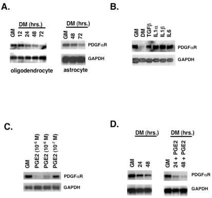
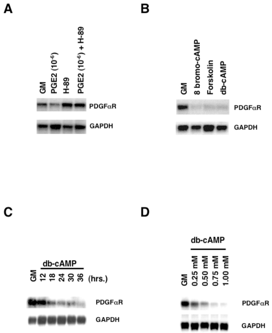
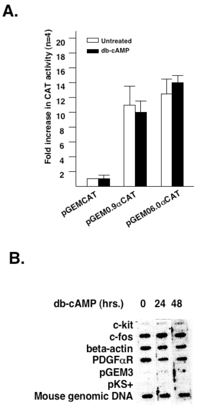
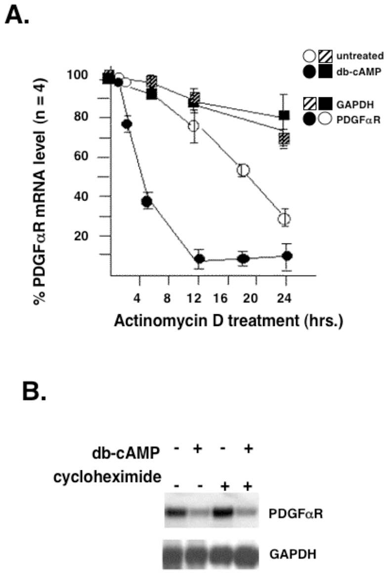
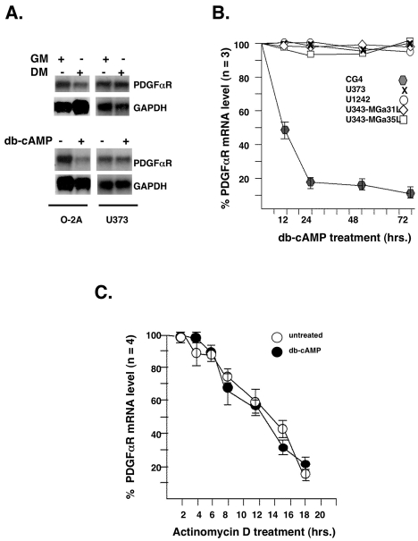
latelet-derived growth factor alpha-receptor (PDGFαR) mediated signaling plays a key role in the development of glial cells of the central nervous system. In vivo and in vitro studies show that PDGFαR is actively expressed in proliferative and motile oligodendrocyte type-2 astrocyte (O-2A) glial progenitor cells. However, PDGFαR expression is barely detectable in mature glial cells. The exact mechanism underlying the loss of PDGFαR expression is unknown. In this study, we employed a rat brain-derived O-2A glial progenitor cell line, CG4 as a culture model to investigate signals capable of inhibiting PDGFαR gene expression. PDGFαR mRNA levels decreased significantly as CG4 cells differentiated into both oligodendrocyte and astrocyte lineages. We showed that inhibition of PDGFαR expression was promoted by prostaglandin E2 via protein kinase A activation. Both cAMP analogs (db-cAMP and 8'bromo-cAMP) and adenylate cyclase activator (forskolin) were potent suppressors of PDGFαR expression in CG4 cells. This inhibitory effect resulted from an increased destabilization of PDGFαR mRNA instead of a decreased PDGFαR gene transcription. Importantly, db-cAMP failed to reduce PDGFαR mRNA levels in several PDGFαR over-expressing human glioma cell lines. Together, these results suggest that cAMP-dependent pathway played a key regulatory role in controlling PDGFαR mRNA levels during normal glial development, and that a breakdown in the cross talk between cAMP and PDGF pathways may underlie the uncontrolled proliferation and immature differentiation state in the glial tumors.

Keywords: PDGF; cyclic AMP; glioma; mRNA turnover.


Figures

Figure 1
Figure 1
Regulation of PDGFαR mRNA levels in O-2A progenitor cells under growth (GM) and differentiation (DM) conditions. (A) CG4 cells were induced to differentiation into oligodendrocytes (left panel) or into astrocytes (right panel) under the condition described in Materials and Methods. Total RNA was prepared from CG4 cells at indicated times of differentiation. (B) Growth factors and chemokines known to promote oligodendrocyte differentiation did not affect PDGFαR mRNA levels in O-2A progenitor cells. CG4 cells grown at 70% confluence in GM (lane 1) either were switched to DM (lane 2) or were treated in GM with TGFβ (10 ng/ml, lane 3), IL-1α (10 ng/ml, lane 4), IL-1β (10 ng/ml, lane 5), and IL-6 (10 ng/ml, lane 6) for 48 hours before harvesting for RNA preparation and analysis. (C) PGE2 suppressed PDGFαR mRNA levels in a concentration-dependent manner. Proliferating CG4 cells in GM were treated with the indicated concentrations of PGE2 for 48 hours. RNA was collected and analyzed. (D) PGE2 accelerated the rate of decline of PDGFαR mRNA levels in differentiating oligodendrocytes. CG4 cells grown in oligodendrocyte-inducing DM were treated with (right panel) or without (left panel) PGE2 (1×10⁻⁶ M) for the time indicated. RNA was collected and analyzed. Northern blot analysis was used to assess the steady state level of PDGFαR mRNA expressed from samples collected under specified conditions. GAPDH mRNA was used as an internal control for normalizing sample loading.
Figure 2
Figure 2
Suppression of PDGFαR gene expression in CG4 cells involved cAMP-dependent protein kinase A pathway. (A) Inhibition of protein kinase A abrogated PGE2-induced decrease in PDGFαR mRNA content. CG4 cells grown in GM were treated without (lanes 1 and 3) or with PGE2 (lanes 2 and 4) in the absence (lanes 1 and 2) and presence (lanes 3 and 4) of 10 μM protein kinase A inhibitor H-89 for 48 hours before harvesting for RNA preparation. (B) Cyclic AMP analogs and forskolin suppressed PDGFαR expression in O-2A progenitor cells. CG4 cells grown in GM were treated without (lane 1) or with 1 mM 8'bromo-cAMP (lane 2), 50 μM forskolin (lane 3), or 1 mM db-cAMP (lane 4) for 48 hours before harvesting for RNA preparation. Time (C) and concentration (D) dependent regulation of PDGFαR gene expression by db-cAMP. For determining dose dependence of db-cAMP on PDGFαR mRNA content, CG4 cells in GM were treated with the indicated concentration of db-cAMP for 24 hours. For determining time dependence, CG4 cells were treated with 1 mM db-cAMP for the indicated times.
Figure 3
Figure 3
Cyclic-AMP treatment did not affect the rate of PDGFαR gene transcription in O-2A progenitor cells as determined by promoter (A) and nuclear run-on (B) analyses. (A) CG4 cells were transiently transfected in duplicates, each with a total of 10 μg DNA containing 8 μg CAT reporter DNA containing without promoter sequence (pGEMCAT) or with PDGFαR promoter sequences (pGEM0.9αCAT and pGEM6.0αCAT) and 2 μg of CMV-lacZ. After transfection, cells were treated with or without 1 mM db-AMP for 48 hours before harvesting for LacZ and CAT assays. LacZ assay was used to correct for variable transfection efficiency. Fold induction in CAT activity was defined as the level of PDGFαR promoter-CAT activity over that of pGEM-CAT promoterless construct measured in the absence of 1 mM db-cAMP. The pGEMCAT activity from untreated cells was given an arbitrary value of 1. Values were calculated based on minimum of three experiments. (B) Nuclei were prepared from CG4 cells with or without treatment of 1 mM db-cAMP for 24 hours. Equal amount of radioactivity corresponding to labeled nascent RNA from each transcription reaction was hybridized to the nylon strip containing indicated DNA templates. The strips were processed under condition described in Materials and methods.
Figure 4
Figure 4
Cyclic-AMP treatment altered the rate of PDGFαR mRNA degradation in O-2A progenitor cells. (A) Effect of db-cAMP on the half-lives of PDGFαR and GADPH mRNAs. Because the db-cAMP effect was not immediate, CG4 cells were pre-treated with 1 mM db-cAMP for 12 hours before the addition of actinomycin -D (5 μg/ml). Total RNA was collected from actinomy-cin-D treated cells at indicated time intervals and analyzed for PDGFαR and GAPDH expression. Radioactive signals were quantified using phosphoimager. The levels of PDGFαR and GAPDH mRNAs in cells treated with or without db-cAMP prior to actinomycin D addition were assigned an arbitrary number of 100%. (B) The effect of cycloheximide on db-cAMP regulated PDGFαR expression in O-2A progenitor cells. CG4 cells grown in GM were treated without (lanes 1 and 3) or with (lanes 2 and 4) 1 mM db-cAMP in the absence (lanes 1 and 2) or presence (lanes 3 and 4) of 5 μg/ml cycloheximide for 18 hours.
Figure 5
Figure 5
Human glioma cells were resistant to cAMP-induced PDGFαR mRNA degradation. (A) Northern blot analysis on the levels of PDGFαR mRNA in CG4 and a human glioma cell line (U373) upon 24-hour treatment with DM (upper panel) or 1 mM db-cAMP (lower panel). (B) Quantitative analysis of the effect of db-cAMP on PDGFαR mRNA levels in CG4 and four human glioma cell lines (U373, U1242, U343-MGa31L, U343-MGa35L) over a period of 72 hours. The levels of PDGFαR mRNA corrected for the GAPDH mRNA are reported as % PGDFαR mRNA at zero time point (untreated). (C) Actinomycin D treatment did not affect the half-life of PDGFαR mRNA in U373 glioma cell line. The experimental condition was the same as described in Figure 4A legend.

Similar articles

References

    1. Bergsten E, Uutela M, Li X, Pietras K, Ostman A, Heldin CH, Alitalo K, Eriksson U. PDGF-D is a specific, protease-activated ligand for the PDGF beta-receptor. Nat Cell Biol. 2001;3:512–516. - PubMed
    1. Bowen Pope DF, Hart CE, Seifert RA. Sera and conditioned media contain different isoforms of platelet-derived growth factor (PDGF) which bind to different class of PDGF receptor. J Biol Chem. 1983;254:2502–2508. - PubMed
    1. Doolittle RF, Hunkapiller MW, Hood LE, Devare SG, Robbins KC, Aaronson SA, Antoniades HN. Simian sarcoma virus oncogene, v-sis, is derived from the gene (or genes) encoding a platelet-derived growth factor. Science. 1983;221:275–277. - PubMed
    1. LaRochelle WJ, Jeffers M, McDonald WF, Chillakuru RA, Giese NA, Lokker NA, Sullivan C, Boldog FL, Yang M, Vernet C, Burgess CE, Fernandes E, Deegler LL, Rittman B, Shimkets J, Shimkets RA, Rothberg JM, Lichenstein HS. PDGF-D, a new protease-activated growth factor. Nat Cell Biol. 2001;3:517–521. - PubMed
    1. Li X, Ponten A, Aase K, Karlsson L, Abramsson A, Uutela M, Backstrom G, Hellstrom M, Bostrom H, Li H, Soriano P, Betsholtz C, Heldin CH, Alitalo K, Ostman A, Eriksson U. PDGF-C is a new protease-activated ligand for the PDGF alpha-receptor. Nat Cell Biol. 2000;2:302–309. - PubMed

LinkOut - more resources