. 2012 May;36(5):748-58.
doi: 10.1111/j.1530-0277.2011.01676.x. Epub 2011 Dec 5.

Ethanol exposure during pregnancy persistently attenuates cranially directed blood flow in the developing fetus: evidence from ultrasound imaging in a murine second trimester equivalent model

Affiliations

Ethanol exposure during pregnancy persistently attenuates cranially directed blood flow in the developing fetus: evidence from ultrasound imaging in a murine second trimester equivalent model

Shameena Bake et al. Alcohol Clin Exp Res. 2012 May.

Abstract

Background: Ethanol (EtOH) consumption during pregnancy can lead to fetal growth retardation, mental retardation, and neurodevelopmental delay. The fetal brain initiates neurogenesis and vasculogenesis during the second trimester, and depends on maternal-fetal circulation for nutrition and growth signals. We used high-resolution in vivo ultrasound imaging to test the hypothesis that EtOH interferes with fetal brain-directed blood flow during this critical developmental period.

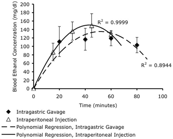
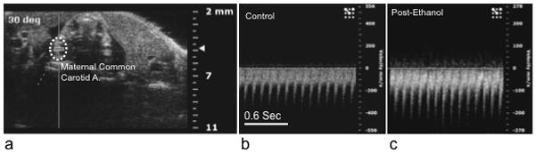
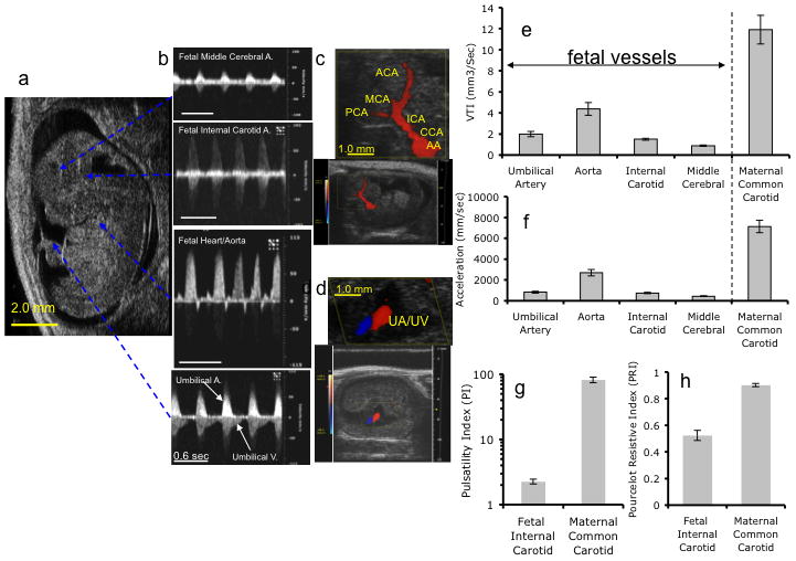
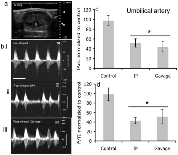
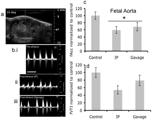
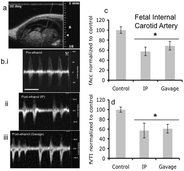
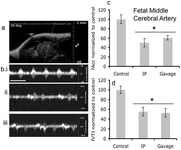
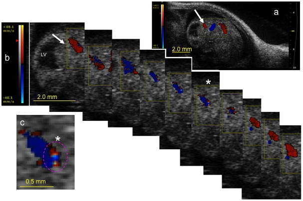
Methods: Pregnant mice were lightly anesthetized on gestational day 12 with an isoflurane/oxygen mixture. We assessed the effect of single and repeated binge-like maternal EtOH exposures at 3 g/kg, administered by intragastric gavage or intraperitoneal injection, on maternal circulation and fetal umbilical, aortic, internal carotid, and middle cerebral arterial circulation.

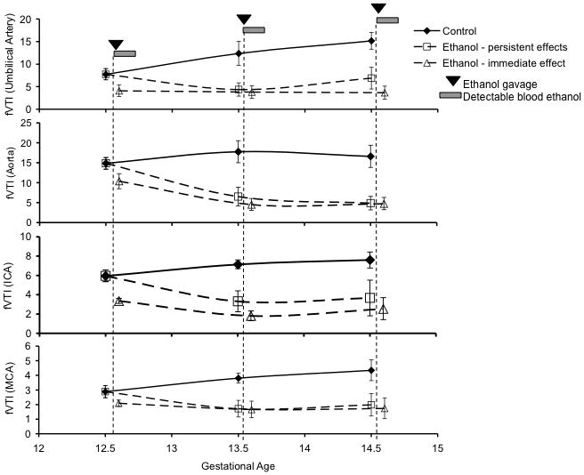
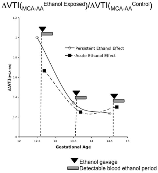
Results: Binge maternal EtOH exposure, regardless of exposure route, significantly reduced fetal arterial blood acceleration and velocity time integral (VTI), from umbilical to cerebral arteries, without a change in fetal heart rate and resistivity indices. Importantly a single maternal binge EtOH exposure induced persistent suppression of fetal arterial VTI for at least 24 hours. Repeated binge episodes resulted in a continuing and persistent suppression of fetal VTI. Qualitative assessments showed that maternal EtOH exposure induced oscillatory, nondirectional blood flow in fetal cerebral arteries. Maternal cardiac and other physiological parameters remained unaltered.

Conclusions: These data show that binge-type maternal EtOH exposure results in rapid and persistent loss of blood flow from the umbilical artery to the fetal brain, potentially compromising nutrition and the maternal/fetal endocrine environment during a critical period for neuron formation and angiogenesis in the maturing brain.


Figures

Figure 1
Figure 1
Blood ethanol concentrations were measured by gas chromatography analysis of mouse tail-vein blood. Intragastric gavage and intraperitoneal ethanol injections resulted in peak blood ethanol concentrations (BEC) of 117mg/dL and 150mg/dL respectively. Data were best fit by second-order polynomial regressions (R2=0.89 and 0.99 respectively), indicating that intragastric gavage resulted in a slightly delayed peak blood concentration of ethanol compared to intraperitoneal injection. There was no statistically significant difference in peak BEC between either routes of administration.
Figure 2
Figure 2
Maternal ethanol exposure (by either intragastric gavage or intraperitoneal injection did not result in a significant change in maternal heart rate or other hemodynamic measures (see results). (a) Sample Doppler image showing angle of insonation and placement of the data acquisition cursor over the maternal common carotid artery. (b and c) show sample sonograms before and after ethanol exposure. Note that the scaling of the ‘Y’ axis (velocity in mm/sec) is different for images b and c, however, there was not a significant difference in maternal carotid artery peak velocity following ethanol exposure. Abbreviations, A; artery. Scale bar (for b and c), 0.6 sec.
Figure 3
Figure 3
Representative fetal color Doppler Images and pulse waveform recordings obtained from control gestational day (GD) 12.5 mice. a–b Image of GD12.5 fetus (a) and corresponding Doppler waveforms obtained from (b, listed from top to bottom) MCA, ICA, Aorta, and UA (Scale bar, 0.6 sec). (c) Color Doppler image and magnified inset (scale bar, 1mm) depict directional cerebro-fugal blood flow (red) from the aortic arch (AA) through the common carotid artery (CCA), the internal carotid artery (ICA), and the anterior (ACA), middle (MCA) and posterior (PCA) cerebral arteries. The gap between the PCA and the ICA represents the probable location of the anastomotic posterior communicating artery. (d) Color Doppler image and magnified inset (scale bar, 1mm) show blood flow through adjacent umbilical artery (UA) and vein (UV). (e,f) Quantitative analysis of velocity time integral (VTI, e) and acceleration (f) measurements made in respective blood vessels in control GD12.5 fetuses and in maternal common carotid artery, prior to maternal ethanol exposure, demonstrating that in the fetus, the greatest flow was observed in the ascending Aorta for both acceleration and VTI. However, fetal Acc, VTI, PI (g) and PRI (h) are all lower than maternal values.
Figure 4
Figure 4
Fluoro-Jade stained sections of the fetal mouse brain show that repeated color and pulse wave Doppler ultrasound scans between GD12.5 and 14.5 do not induce cell death. (a,b) Composite photomicrographs (yellow lines show boundaries of individual images stitched together into a composite) showing saggital brain sections at the level of the lateral ventricle from control littermates (a, unexposed to ultrasound) and ultrasound (b, both color and pulse wave Doppler)-exposed fetuses showed no evidence for cell death. (c–e) Endothelin-1 induced stroke in contrast resulted in significant cell death in the striatum (c, green cellular staining) that is localized both to cells in the vicinity of blood vessels (d) and to neurons (e). Scale bar, 100um.
Figure 5
Figure 5
Fetal UA Doppler ultrasound measurements. (a) Doppler ultrasound image of area where waveform analysis was acquired. (b) Representative Doppler ultrasound image of fetal umbilical artery waveform before alcohol administration and after alcohol treatment via intraperitoneal (IP) injection and Gavage routes. (c,d) Maternal ethanol exposure by gavage or IP resulted in a significant decrease in acceleration and VTI (asterisk indicates statistically significant difference from baseline control). No difference was observed based on route of administration. Scale bar, 0.6 sec.
Figure 6
Figure 6
Doppler ultrasound measurement of fetal Aorta. (a) Doppler ultrasound image of fetal Aorta indicating area of analysis. (b) Doppler ultrasound waveform of fetal Aorta before ethanol treatment and after both IP and Gavage routes of administration. (c,d) Blood flow as measured by Acceleration was significantly reduced following maternal ethanol exposure (asterisk indicates statistically significant difference from baseline control), however, VTI, though exhibiting a declining trend, was not significantly decreased. No difference was observed between routes of administration. Scale bar, 0.6 sec.
Figure 7
Figure 7
Doppler ultrasound measurements of fetal Internal Carotid Artery. (a) Fetal Doppler ultrasound image showing position marked for analysis of Internal Carotid Artery. (b) Representative Doppler ultrasound waveforms of Internal Carotid Artery before ethanol treatment and after ethanol treatment via IP and gavage routes of administration. (c,d) Maternal ethanol exposure induced a significant decline in both acceleration and VTI, with no difference observed between routes of administration (asterisk indicates statistically significant difference from baseline control). Scale bar, 0.6 sec.
Figure 8
Figure 8
Doppler ultrasound measurements of fetal Middle Cerebral Artery. (a) Fetal Doppler ultrasound image of middle cerebral artery. (b) Doppler ultrasound recordings of the middle cerebral artery before ethanol treatment and after via intraperitoneal and Gavage routes of treatment. (c,d) Doppler ultrasound measurements showed that maternal ethanol exposure induced a significant reduction in acceleration and VTI in the middle cerebral artery, irrespective of the route of administration (asterisk indicates statistically significant difference from baseline control). Scale bar, 0.6 sec.
Figure 9
Figure 9
Color Doppler image series document a loss of directional blood flow in the fetal posterior cerebral artery following maternal ethanol exposure. Red color overlay indicates blood flow towards the ultrasonic transducer, whereas a blue overlay indicates flow directed away from the transducer. (a) Low magnification image showing location of the active color Doppler acquisition window (yellow box). White arrow points to the location of the posterior cerebral artery and corresponds to the image sequence in ‘b’. (b) Sequential images acquired over a single cardiac cycle show alternating blood flow towards (red) and away (blue) from the ultrasonic transducer within the same arterial cross-section. Color scale bar (left) indicates velocity (mm/sec). (c) Magnified frame from (b, asterisk) shows the simultaneous presence of adjacent red and blue pixels within the same luminal cross-section, suggesting locally turbulent blood flow. Oscillatory blood flow within the posterior cerebral artery suggests that maternal ethanol exposure can lead to fetal distress. Scale bars; a and b = 2.0mm, c = 0.5mm. Abbreviations, LV, lateral ventricle.
Figure 10
Figure 10
Repeated maternal ethanol exposure results in persistent suppression of fVTI in fetal blood vessels. Pregnant mice were exposed to ethanol (3g/kg) once a day by intragastric gavage for three consecutive days (GD12.5–14.5) and were imaged daily before and following maternal ethanol exposure to detect changes in fetal blood flow. Shown from top to bottom are graphical representations of changes in fVTI in fetal umbilical artery, aorta, internal carotid artery and middle cerebral artery. A persistent fetal effect is defined as one that can be observed 24 hours after an episode of maternal ethanol exposure (i.e., an effect observed at GD13.5 and 14.5 due to exposures on GD12.5 and 13.5), while the acute effect is defined as the additional effect of a maternal binge-like exposure episode on that day, in the period immediately preceding the ultrasound scan. In all vessels, we observed a main effect of ethanol exposure, but no interaction between gestational age and ethanol exposure on fVTI. Visual inspection of the data show that maternal ethanol exposure on GD12.5 induced a persistent decrease in fVTI 24 hours later, and this decrease in fVTI persisted with repeated maternal ethanol exposure, though the magnitude of the decline did not increase with subsequent exposures.
Figure 11
Figure 11
Analysis of the ratio of the decrease in fVTI from the fetal aorta to the fetal middle cerebral artery before and following maternal ethanol exposure (ΔΔfVTI(MCA-AA)) reveals evidence for an adaptive fetal ‘brain-sparing’ response to maternal ethanol exposure. Repeated ethanol exposure resulted in both an acute and a persistent (i.e., an effect observed at GD13.5 and 14.5 due to exposures on GD12.5 and 13.5) drop in the ΔΔfVTI(MCA-AA) ratio suggesting that the decline in fVTI at the MCA is less that predicted from the drop in fVTI at the aorta.

Similar articles

Cited by

References

    1. Abel EL. Prenatal effects of alcohol. Drug Alcohol Depend. 1984;14(1):1–10. - PubMed
    1. Abel EL. An update on incidence of FAS: FAS is not an equal opportunity birth defect. Neurotoxicol Teratol. 1995;17(4):437–43. - PubMed
    1. Bates DO. Vascular endothelial growth factors and vascular permeability. Cardiovasc Res. 2010;87(2):262–71. - PMC - PubMed
    1. Bayer S, Altman J, Russo R, Zhang X. Timetables of neurogenesis in the human brain based on experimentally determined patterns in the rat. Neuro Toxicology. 1993;14(1):83–144. - PubMed
    1. Bearer CF. L1 cell adhesion molecule signal cascades: targets for ethanol developmental neurotoxicity. Neurotoxicology. 2001;22(5):625–33. - PubMed

Publication types

MeSH terms