. 2012 May;166(2):702-20.
doi: 10.1111/j.1476-5381.2011.01802.x.

17β-Oestradiol inhibits doxorubicin-induced apoptosis via block of the volume-sensitive Cl(-) current in rabbit articular chondrocytes

Affiliations

17β-Oestradiol inhibits doxorubicin-induced apoptosis via block of the volume-sensitive Cl(-) current in rabbit articular chondrocytes

Kousuke Kumagai et al. Br J Pharmacol. 2012 May.

Abstract

BACKGROUND AND PURPOSE Chondrocyte apoptosis contributes to disruption of cartilage integrity in osteoarthritis. Recent evidence suggested that the volume-sensitive organic osmolyte/anion channel [volume-sensitive (outwardly rectifying) Cl(-) current (I(Cl,vol) )] plays a functional role in the development of cell shrinkage associated with apoptosis (apoptotic volume decrease) in several cell types. In this study, we investigated the cellular effects of 17β-oestradiol on doxorubicin-induced apoptotic responses in rabbit articular chondrocytes. EXPERIMENTAL APPROACH Whole-cell membrane currents and cross-sectional area were measured from chondrocytes using a patch-clamp method and microscopic cell imaging, respectively. Caspase-3/7 activity was assayed as an index of apoptosis. KEY RESULTS Addition of doxorubicin (1 µM) to isosmotic bath solution rapidly activated the Cl(-) current with properties similar to those of I(Cl,vol) in chondrocytes. Doxorubicin also gradually decreased the cross-sectional area of chondrocytes, followed by enhanced caspase-3/7 activity; both of these responses were totally abolished by the I(Cl,vol) blocker DCPIB (20 µM). Pretreatment of chondrocytes with 17β-oestradiol (1 nM) for short (approximately 10 min) and long (24 h) periods almost completely prevented the doxorubicin-induced activation of I(Cl,vol) and subsequent elevation of caspase-3/7 activity. These effects of 17β-oestradiol were significantly attenuated by the oestrogen receptor blocker ICI 182780 (10 µM), as well as the phosphatidyl inositol-3-kinase (PI3K) inhibitors wortmannin (100 nM) and LY294002 (20 µM). Testosterone (10 nM) had no effect on the doxorubicin-induced Cl(-) current. CONCLUSIONS AND IMPLICATIONS 17β-Oestradiol prevents the doxorubicin-induced cell shrinkage mediated through activation of I(Cl,vol) and subsequent induction of apoptosis signals, through a membrane receptor-dependent PI3K pathway in rabbit articular chondrocytes.


Figures

Figure 1
Figure 1
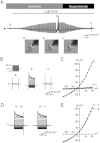
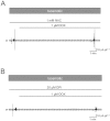
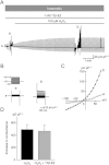
Whole-cell membrane currents evoked by doxorubicin in rabbit articular chondrocytes. (A) Time course of changes in whole-cell currents recorded from a chondrocyte, initially exposed to doxorubicin (DOX, 1 µM) in isosmotic solution (360 mosmol·L−1) and then to hyperosmotic solution (430 mosmol·L−1) during the continued presence of doxorubicin, as denoted above the current traces. The vertical deflections of current trace reflect the imposition of either voltage ramps or square steps. Microscopic images of whole-cell clamped chondrocyte taken before (a) and during (b) exposure to doxorubicin in isosmotic solution and after switching to hyperosmotic solution in the presence of doxorubicin (c). The calibration bar represents 10 µm. The diameter of the chondrocyte was measured to be 14.1 (a), 14.1 (b) and 12.7 µm (c), respectively, in each condition. (B) Superimposed current traces in response to 200 ms square-steps applied from a holding potential of −30 mV to test potentials of +80 through −100 mV in 10 mV steps, at the time points identified by letters (a, b and c) in (A). (C) IV relationships of whole-cell currents recorded at each time point (a, b and c) shown in (B). (D) Membrane currents activated by doxorubicin in isosmotic solution (b–a) and those inhibited by hyperosmotic solutions in the presence of doxorubicin (b–c), obtained by digital subtraction of current traces, as indicated. (E) IV relationships for the doxorubicin-induced currents (b–a) and hyperosmotic solution-inhibited currents (b–c), shown in (D).
Figure 2
Figure 2
Electrophysiological and pharmacological properties of ICl,Dox. (A) The relationship between the reversal potential (Erev) of the doxorubicin (1 µM)-activated current and extracellular Cl concentrations ([Cl]o), obtained with an internal solution containing 54 mM Cl. Data points represent mean ± SEM (n = 5). The straight line represents a linear regression fit to the data points. (B) IV relationship of ICl,Dox in the presence of an equivalent concentration of Cl inside and outside the cell ([Cl]i = [Cl]o = 54 mM). The inset shows the changes in membrane current during exposure to 1 µM doxorubicin in isosmotic conditions in equivalent Cl (54 mM). (C) and (D) Chondrocyte was initially exposed to 1 µM doxorubicin, and then to 20 µM DCPIB (C) or 30 µM arachidonic acid (AA, D) during the continued presence of 1 µM doxorubicin, as indicated. Whole-cell current was recorded during repetitive imposition of voltage ramps and in part by square voltage-steps (B–D).
Figure 3
Figure 3
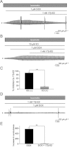
Inhibitory action of 17β-oestradiol on ICl,Dox mediated via its plasma membrane receptor. (A) Inhibition of doxorubicin (DOX, 1 µM)-induced activation of ICl,Dox by subsequent application of 17β-oestradiol (17β-E2, 1 nM). (B) Effect of pre-exposure to ICI 182780 (ICI, 10 µM) on 17β-oestradiol-induced inhibition of ICl,Dox. (C) % inhibition of ICl,Dox by 17β-oestradiol in the absence and presence of ICI 182780 (10 µM). **P < 0.01 (Student's unpaired t-test). (D) Effect of pre-exposure to 17β-oestradiol (1 nM) on the activation of ICl,Dox. ICI 182780 (B) or 17β-oestradiol (D) was added approximately 10 min before the application of doxorubicin (DOX, 1 µM) and was present throughout the experiments. (E) Slope conductance of ICl,vol activated by doxorubicin (1 µM) without and with pre-exposure to 17β-oestradiol. **P < 0.01 (Student's unpaired t-test).
Figure 4
Figure 4
Lack of effects of 17β-oestradiol on the ICl,vol activation by cell swelling. (A) Time course of changes in whole-cell current recorded from a chondrocyte during switching from isosmotic to hyposmotic external solution in the continuous presence of 17β-oestradiol (1 nM). 17β-oestradiol was added to the bath (isosmotic solution) approximately 10 min before switching to hyposmotic solution. Inset shows microscopic images of whole-cell clamped chondrocyte taken before (a) and after (b) switching to hyposmotic solution, and diameter in each condition was calculated to be 13.3 and 16.1 µm, respectively. (B) Superimposed current traces in response to 200-ms square steps applied from a holding potential of −30 mV to potentials of +80 through −100 mV in 10 mV steps, in isosmotic (a) and hyposmotic (b) solutions. (C) IV relationships of membrane currents in isosmotic (a) and hyposmotic (b) solutions, shown in (B). (D) Increased conductance of ICl,vol recorded without and with 17β-oestradiol pretreatment. There was no significant difference in the degree of ICl,vol activation between these two groups (561.0 ± 100.6 pS·pF−1, n = 9, N = 5 versus 551.9 ± 91.5 pS·pF−1, n = 8, N = 5; P = 0.50). (E) Time course of changes in cross-sectional area during hyposmotic challenge without (Control) and with 17β-oestradiol (1 nM) pretreatment. Data points represent means and SEM of 15 (N = 3) and 18 (N = 3) chondrocytes without and with 17β-oestradiol pretreatment, respectively.
Figure 5
Figure 5
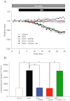
Doxorubicin-induced AVD and caspase-3/7 activity in chondrocytes. (A) Time course of changes in cross-sectional area of cell (chondrocyte) image in the absence (Control) and presence of doxorubicin (1 µM) applied without or with DCPIB (20 µM), 17β-oestradiol (1 nM) and/or ICI 182780 (10 µM), as indicated. All these compounds were added to the isosmotic solution at time 0. (B) Caspase-3/7 activity, measured from chondrocytes after 24 h exposure to doxorubicin (1 µM) without or with DCPIB (20 µM), 17β-oestradiol (1 nM) and/or ICI 182780 (10 µM). Asterisks represent P values according to Newman–Keuls multiple means comparison test (**P < 0.01).
Figure 6
Figure 6
Involvement of reactive oxygen species in the activation of ICl,Dox. (A) and (B) Abolishment of ICl,Dox activation by pretreatment with the ROS scavenger N-acetyl-cysteine (NAC) at 1 mM (A) or the general inhibitor of NAD(P)H oxidase diphenylene-iodonium chloride (DPI) at 20 µM (B). N-acetyl-cysteine (A) or diphenylene-iodonium chloride (B) was added approximately 10 min before the application of doxorubicin (DOX, 1 µM) and was present throughout the experiments.
Figure 7
Figure 7
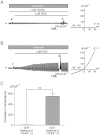
Lack of effects of 17β-oestradiol on ICl,vol activation induced by hydrogen peroxide. (A) Time course of changes in whole-cell current during exposure to hydrogen peroxide (100 µM) in isosmotic solution in a chondrocyte pretreated with 17β-oestradiol (1 nM). 17β-Oestradiol was added to isosmotic solution approximately 10 min before exposure to hydrogen peroxide and was present throughout the experiments. (B) Superimposed current traces in response to 200-ms square steps applied from a holding potential of −30 mV to test potentials of +80 through −100 mV in 10 mV steps before (a) and during (b) exposure to hydrogen peroxide (100 µM) in the presence of 17β-oestradiol (1 nM). (C) IV relationships for whole-cell currents recorded at each time points (a and b) in (B). (D) Maximal increase in the slope conductance for ICl,Dox without and with pretreatment of 17β-oestradiol (1 nM). There was no significant difference between the control (n = 4, N = 4) and 17β-oestradiol (n = 4, N = 4) groups (P = 0.27).
Figure 8
Figure 8
Involvement of PI3-kinase activity in the inhibitory effect of 17β-oestradiol on of ICl,Dox. (A) and (B) The effect of pretreatment with 100 nM wortmannin (Wort, A) or 20 µM LY294002 (LY, B) on the inhibitory effect of 17β-oestradiol (1 nM) on ICl,Dox. Various test compounds were added to isosmotic bath solutions, as indicated. (C) Percentage inhibition of ICl,Dox by 17β-oestradiol without and with pretreatment with wortmannin and LY294002. (D) Caspase-3/7 activity was measured after 24 h treatment without (Control) and with doxorubicin in the absence and presence of 17β-oestradiol, wortmannin (100 nM) and/or LY294002 (20 µM). Asterisks represent P values according to Newman–Keuls multiple means comparison test (*P < 0.05, **P < 0.01).
Figure 9
Figure 9
Effect of a longer pre-incubation with 17β-oestradiol on the activation of ICl,Dox in chondrocytes. (A) Abolishment of ICl,Dox activation in chondrocytes pre-incubated with 17β-oestradiol for 24 h without washing out (17β-oestradiol was present throughout the experiment). (B) Activation of ICl,Dox in chondrocytes when 17β-oestradiol was washed out for 1 h after 24 h of pre-incubation. Insets (right) in (A and B) show the IV relationship of difference current representing the doxorubicin (1 µM)-evoked current. (C) Increase in conductance of ICl,Dox in chondrocytes pre-incubated with 17β-oestradiol for 24 h without (6.6 ± 1.7 pS·pF−1, n = 5, N = 3) and with (552.5 ± 44.3 pS·pF−1, n = 5, N = 3) washing out the hormone for 1 h. **P < 0.01 between the two groups (Student's unpaired t-test).
Figure 10
Figure 10
Effect of longer pre-incubation with 17β-oestradiol on the doxorubicin-induced changes in cell size and caspase-3/7 activity. (A) Changes in cell size (as assessed by relative area) during exposure to doxorubicin (1 µM) in chondrocytes pre-incubated with 17β-oestradiol for 24 h without washing out the hormone (17β-oestradiol was present throughout the experiment) or with washing out for 1 h (17β-oestradiol was absent during the measurement). (B) Caspase-3/7 activity in chondrocytes pre-incubated with 17β-oestradiol for 24 h without and with washing out the hormone for 1 h. **P < 0.01 between the two groups (Student's unpaired t-test).
Figure 11
Figure 11
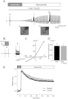
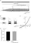
Lack of inhibitory effect of testosterone on ICl,Dox. (A) Time course of changes in whole-cell current recorded from a chondrocyte that was initially exposed to doxorubicin (DOX, 1 µM) and subsequently to doxorubicin plus testosterone (10 nM) in isosmotic solution. (B) Superimposed current traces in response to 200 ms square-steps applied from a holding potential of −30 mV to test potentials of +80 through −100 mV in 10 mV steps, at the time points identified by letters (a, b and c) in (A). (C) IV relationships of whole-cell currents recorded at each time point (a, b and c) shown in (B). (D) Increased conductance of ICl,Dox measured at a steady-state activation (DOX) and 10 min after subsequent addition of 10 nM testosterone (DOX + Testosterone). There was no significant difference in the degree of ICl,Dox activation between these two conditions (580.9 ± 45.4 pS·pF−1 versus 588.4 ± 32.9 pS·pF−1, n = 5, N = 3; P = 0.84).

Similar articles

Cited by

References

    1. Adams CS, Shapiro IM. The fate of the terminally differentiated chondrocyte: evidence for microenvironmental regulation of chondrocyte apoptosis. Crit Rev Oral Biol Med. 2002;13:465–473. - PubMed
    1. Alexander SPH, Mathie A, Peters JA. Guide to Receptors and Channels (GRAC), 5th Edition. Br J Pharmacol. 2011;164(Suppl. 1):S1–S324. - PMC - PubMed
    1. Almeida M, Martin-Millan M, Ambrogini E, Bradsher R, 3rd, Han L, Chen X-D, et al. Estrogens attenuate oxidative stress and the differentiation and apoptosis of osteoblasts by DNA-binding-independent actions of the ERα. J Bone Miner Res. 2010;25:769–781. - PMC - PubMed
    1. d'Anglemont de Tassigny A, Souktani R, Henry P, Ghaleh B, Berdeaux A. Volume-sensitive chloride channels (ICl,vol) mediate doxorubicin-induced apoptosis through apoptotic volume decrease in cardiomyocytes. Fundam Clin Pharmacol. 2004;18:531–538. - PubMed
    1. Bénitah JP, Gomez AM, Bailly P, Da Ponte JP, Berson G, Delgado C, et al. Heterogeneity of the early outward current in ventricular cells isolated from normal and hypertrophied rat hearts. J Physiol. 1993;469:111–138. - PMC - PubMed

Publication types