

Localization of acidic phospholipid cardiolipin and DnaA in mycobacteria
- PMID: 22142462
- PMCID: PMC3249017
- DOI: 10.1016/j.tube.2011.10.025
Localization of acidic phospholipid cardiolipin and DnaA in mycobacteria
Abstract
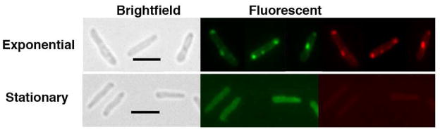
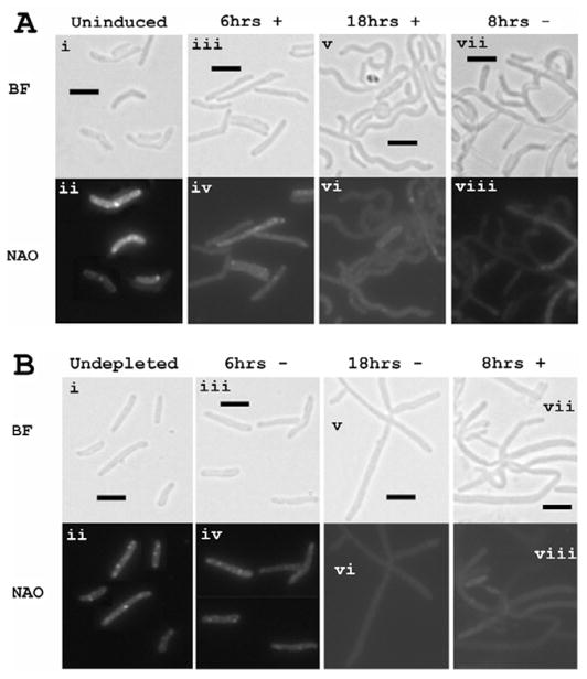
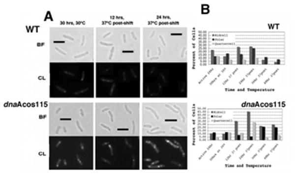
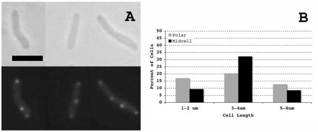
Acidic phospholipids such as cardiolipin (CL) have been shown to modulate Mycobacterium tuberculosis (Mtb) DnaA interactions with ATP. In the present study, using nonyl acridine orange fluorescent dye we localized CL-enriched regions to midcell septa and poles of actively dividing cells. We also found that CL-enriched regions were not visualized in cells defective for septa formation as a consequence of altered FtsZ levels. Using Mtb cultures synchronized for DNA replication we show that CL localization could be used as a marker for cell division and cell cycle progression. Finally, we show that the localization pattern of the DnaA-green fluorescent fusion protein is similar to CL. Our results suggest that DnaA colocalizes with CL during cell cycle progression.
Copyright © 2011 Elsevier Ltd. All rights reserved.
Conflict of interest statement
Competing interests: The authors have no conflicts of interest to declare.
Figures
Similar articles
-
Replacement of Mycobacterium smegmatis dnaA gene by Mycobacterium tuberculosis homolog results in temperature sensitivity.Tuberculosis (Edinb). 2011 Dec;91 Suppl 1:S136-41. doi: 10.1016/j.tube.2011.10.023. Epub 2011 Nov 22. Tuberculosis (Edinb). 2011. PMID: 22112933 Free PMC article.
-
Septal localization of the Mycobacterium tuberculosis MtrB sensor kinase promotes MtrA regulon expression.J Biol Chem. 2012 Jul 6;287(28):23887-99. doi: 10.1074/jbc.M112.346544. Epub 2012 May 20. J Biol Chem. 2012. PMID: 22610443 Free PMC article.
-
Overproduction and localization of Mycobacterium tuberculosis ParA and ParB proteins.Tuberculosis (Edinb). 2009 Dec;89 Suppl 1(Suppl 1):S65-9. doi: 10.1016/S1472-9792(09)70015-0. Tuberculosis (Edinb). 2009. PMID: 20006309 Free PMC article.
-
Crosstalk between DnaA protein, the initiator of Escherichia coli chromosomal replication, and acidic phospholipids present in bacterial membranes.Int J Mol Sci. 2013 Apr 17;14(4):8517-37. doi: 10.3390/ijms14048517. Int J Mol Sci. 2013. PMID: 23595001 Free PMC article. Review.
-
Perspective: challenges and opportunities for the study of cardiolipin, a key player in bacterial cell structure and function.Curr Genet. 2018 Aug;64(4):795-798. doi: 10.1007/s00294-018-0811-2. Epub 2018 Feb 9. Curr Genet. 2018. PMID: 29427078 Review.
Cited by
-
Chromosome Replication in Escherichia coli: Life on the Scales.Life (Basel). 2012 Oct 29;2(4):286-312. doi: 10.3390/life2040286. Life (Basel). 2012. PMID: 25371267 Free PMC article.
-
Cardiolipin signaling mechanisms: collapse of asymmetry and oxidation.Antioxid Redox Signal. 2015 Jun 20;22(18):1667-80. doi: 10.1089/ars.2014.6219. Epub 2015 Mar 31. Antioxid Redox Signal. 2015. PMID: 25566681 Free PMC article. Review.
-
Cardiolipin and mitochondrial cristae organization.Biochim Biophys Acta Biomembr. 2017 Jun;1859(6):1156-1163. doi: 10.1016/j.bbamem.2017.03.013. Epub 2017 Mar 20. Biochim Biophys Acta Biomembr. 2017. PMID: 28336315 Free PMC article. Review.
-
The membrane: transertion as an organizing principle in membrane heterogeneity.Front Microbiol. 2015 Jun 12;6:572. doi: 10.3389/fmicb.2015.00572. eCollection 2015. Front Microbiol. 2015. PMID: 26124753 Free PMC article. Review.
-
Mycobacterium tuberculosis proteins involved in mycolic acid synthesis and transport localize dynamically to the old growing pole and septum.PLoS One. 2014 May 9;9(5):e97148. doi: 10.1371/journal.pone.0097148. eCollection 2014. PLoS One. 2014. PMID: 24817274 Free PMC article.
References
Publication types
MeSH terms
Substances
Grants and funding
LinkOut - more resources
Full Text Sources
