Review
. 2011 Dec;21(6):785-91.
doi: 10.1016/j.sbi.2011.07.006.

Regulation of protein kinases by lipids

Affiliations
Review

Regulation of protein kinases by lipids

Thomas A Leonard et al. Curr Opin Struct Biol. 2011 Dec.

Abstract

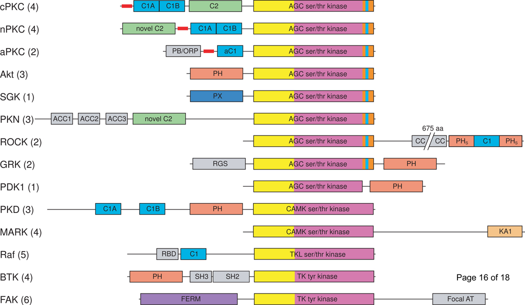
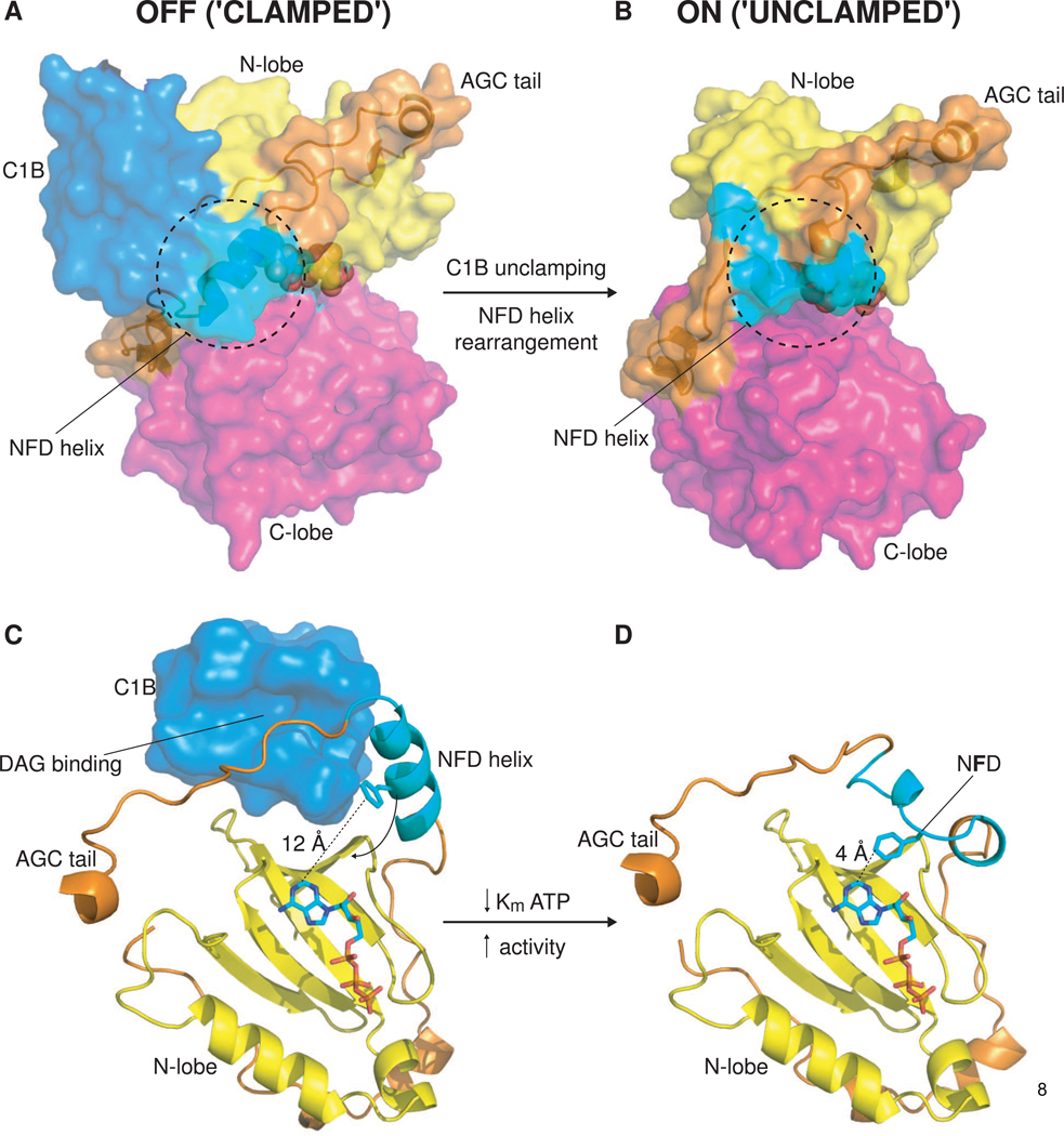
Membranes are sites of intense signaling activity within the cell, serving as dynamic scaffolds for the recruitment of signaling molecules and their substrates. The specific and reversible localization of these signaling molecules to membranes is critical for the appropriate activation of downstream signaling pathways. Phospholipid-binding domains, including C1, C2, PH, and PX domains, play critical roles in the membrane targeting of protein kinases. Recent structural studies have identified a new membrane association domain, the Kinase Associated 1 (KA1) domain, which targets a number of yeast and mammalian protein kinases to membranes containing acidic phospholipids. Despite an abundance of localization studies on lipid-binding proteins and structural studies of the isolated lipid-binding domains, the question of how membrane binding is coupled to the activation of the kinase catalytic domain has been virtually untouched. Recently, structural studies on protein kinase C (PKC) have provided some of the first structural insights into the allosteric regulation of protein kinases by lipid second messengers.


Figures

Figure 1
Figure 1
Domain composition of the major protein kinase families containing lipid-binding domains. Lipid-binding domains are: C1 domain (light blue), C2 domain (green), PH domain (salmon), PX domain (dark blue), KA1 domain (pale orange), FERM domain (purple). The kinase domains are shown in yellow (N-lobe) and magenta (C-lobe); the AGC kinase family C-terminal tail is orange with the NFD motif highlighted in cyan. Pseudosubstrate segments are represented as thick red lines. Additional domains are shown in gray: Bem1 (PB/ORP), antiparallel coiled coil (ACC), coiled coil (CC), regulator of G-protein signaling (RGS), Ras binding domain (RBD), Src homology 2 (SH2). cPKC, conventional protein kinase C; nPKC, novel protein kinase C; aPKC, atypical protein kinase C; SGK, serum/glucocorticoid regulated kinase; PKN, protein kinase N; ROCK, Rho-activated kinase; GRK, Gprotein coupled receptor kinase; PDK1, phosphoinositide-dependent kinase 1; PKD, protein kinase D; MARK, microtubule affinity regulating kinase; focal AT, focal adhesion targeting. Numbers in parentheses indicate the number of mammalian kinases in each family.
Figure 2
Figure 2
Allosteric activation of PKC by DAG binding to the C1B domain. (A) ‘Clamped’ conformation of PKCβII. Surface representation of PKCβII, illustrating the interface between the lipid-binding C1B domain (light blue) and the kinase domain (yellow/magenta). A novel helix containing the conserved NFD motif found in the Cterminal tail of AGC kinases packs against the C1B domain, while residues preceding the novel helix occupy the DAG binding site of the C1B domain. ATP is represented with spheres. (B) ‘Unclamped’ conformation of PKCι. Surface representation of PKCι, illustrating the conformational change that occurs in the NFD helix upon DAG/membrane binding by the C1B domain. (C) View of the DAG-binding cleft of the C1B domain and the nucleotide-binding site (sandwiched between the N-lobe (yellow) and C-lobe (not shown) of the kinase domain). The lipid-binding site is inaccessible in this conformation and the critical phenylalanine of the NFD motif is sequestered 12 Å away from the nucleotide. (D) View of the active conformation of PKCι in which the NFD helix has undergone a conformational rearrangement in the absence of the C1 domain. The central phenylalanine of the NFD motif is brought into close (4 Å) contact with the adenine ring of ATP. Contacts between the phenylalanine side chain and the adenine moiety lower the Km for ATP, thereby increasing the catalytic rate, Kcat, of the enzyme.

References

    1. Huse M, Kuriyan J. The conformational plasticity of protein kinases. Cell. 2002;109:275–282. - PubMed
    1. Pellicena P, Kuriyan J. Protein-protein interactions in the allosteric regulation of protein kinases. Curr Opin Struct Biol. 2006;16:702–709. - PubMed
    1. Lemmon MA. Membrane recognition by phospholipid-binding domains. Nat Rev Mol Cell Biol. 2008;9:99–111. - PubMed
    1. Moravcevic K, Mendrola JM, Schmitz KR, Wang YH, Slochower D, Janmey PA, Lemmon MA. Kinase associated-1 domains drive MARK/PAR1 kinases to membrane targets by binding acidic phospholipids. Cell. 2010;143:966–977. •• This work identifies a new lipid-binding domain found in protein kinases and highlights the importance of coincidence detection in signaling at the membrane.

    1. Fang Y, Vilella-Bach M, Bachmann R, Flanigan A, Chen J. Phosphatidic acid-mediated mitogenic activation of mTOR signaling. Science. 2001;294:1942–1945. - PubMed

Publication types

LinkOut - more resources