

Nanofabrication for the analysis and manipulation of membranes
- PMID: 22143598
- PMCID: PMC4351669
- DOI: 10.1007/s10439-011-0479-y
Nanofabrication for the analysis and manipulation of membranes
Abstract
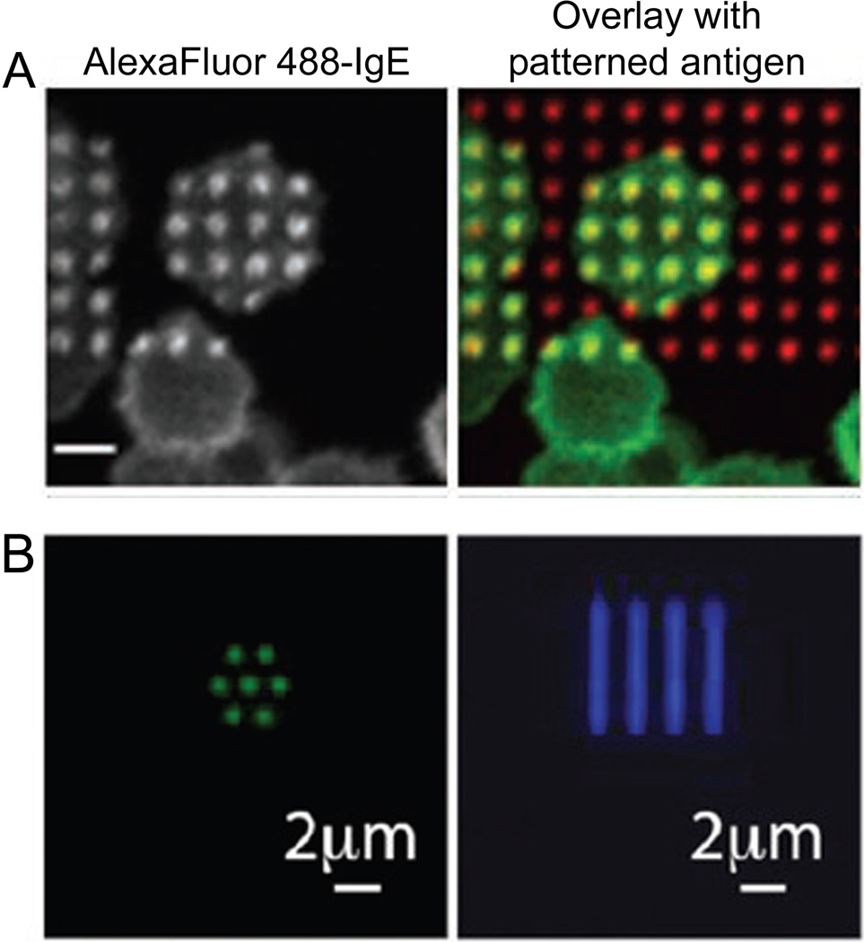
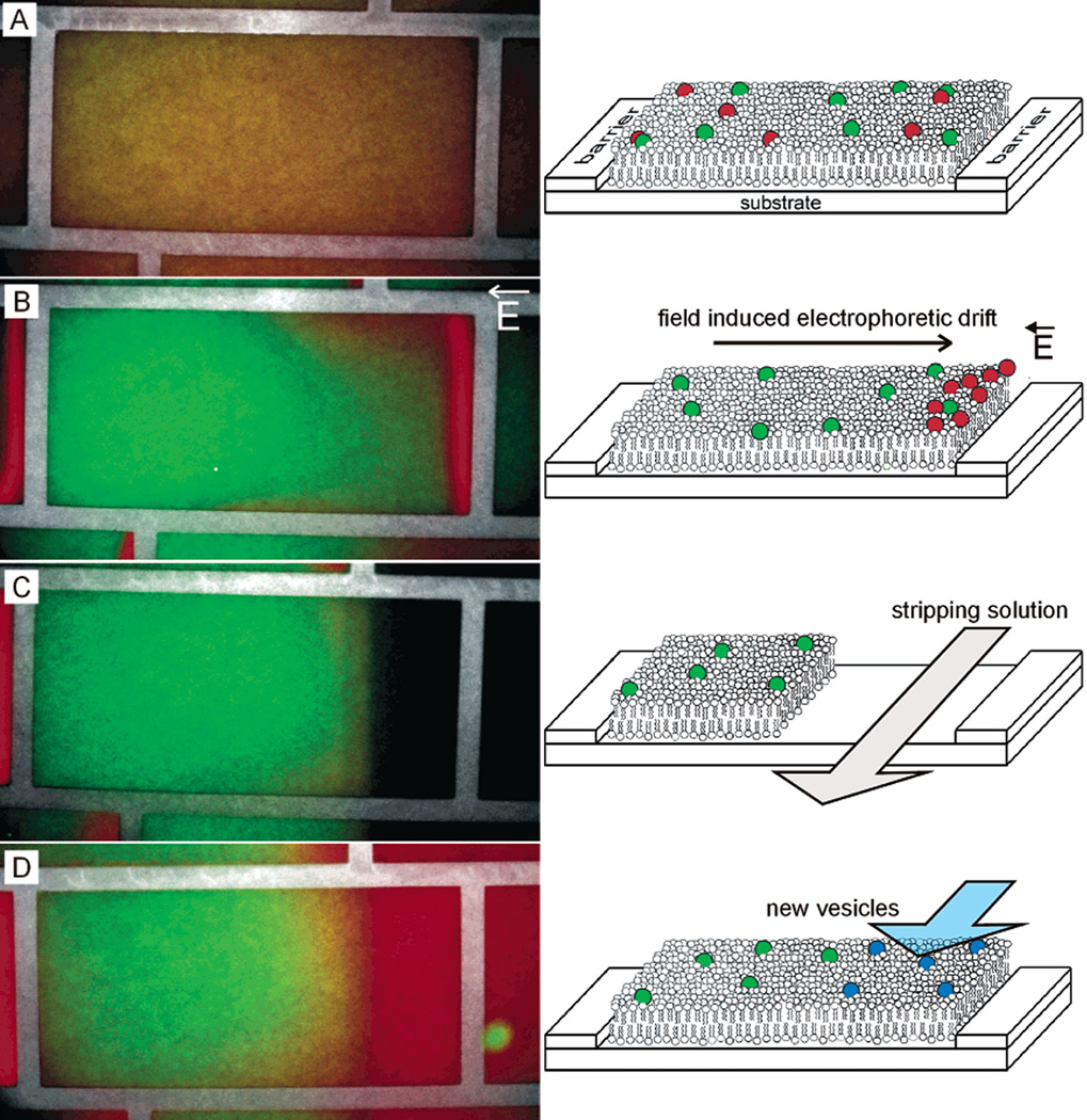
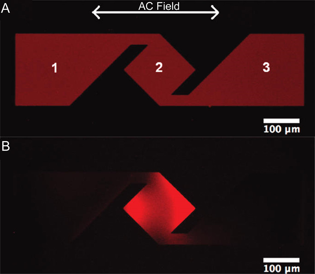
Recent advancements and applications of nanofabrication have enabled the characterization and control of biological membranes at submicron scales. This review focuses on the application of nanofabrication towards the nanoscale observing, patterning, sorting, and concentrating membrane components. Membranes on living cells are a necessary component of many fundamental cellular processes that naturally incorporate nanoscale rearrangement of the membrane lipids and proteins. Nanofabrication has advanced these understandings, for example, by providing 30 nm resolution of membrane proteins with metal-enhanced fluorescence at the tip of a scanning probe on fixed cells. Naturally diffusing single molecules at high concentrations on live cells have been observed at 60 nm resolution by confining the fluorescence excitation light through nanoscale metallic apertures. The lateral reorganization on the plasma membrane during membrane-mediated signaling processes has been examined in response to nanoscale variations in the patterning and mobility of the signal-triggering molecules. Further, membrane components have been separated, concentrated, and extracted through on-chip electrophoretic and microfluidic methods. Nanofabrication provides numerous methods for examining and manipulating membranes for both greater understandings of membrane processes as well as for the application of membranes to other biophysical methods.
Figures
Similar articles
-
Microchip electrophoresis of DNA following preconcentration at photopatterned gel membranes.Electrophoresis. 2012 Apr;33(8):1236-46. doi: 10.1002/elps.201100675. Electrophoresis. 2012. PMID: 22589100
-
Integration of nanoporous membranes for sample filtration/preconcentration in microchip electrophoresis.Electrophoresis. 2006 Dec;27(24):4927-34. doi: 10.1002/elps.200600252. Electrophoresis. 2006. PMID: 17117457
-
Ion-permeable membrane for on-chip preconcentration and separation of cancer marker proteins.Electrophoresis. 2011 May;32(10):1133-40. doi: 10.1002/elps.201000698. Electrophoresis. 2011. PMID: 21544838 Free PMC article.
-
Integrating amperometric detection with electrophoresis microchip devices for biochemical assays: recent developments.Talanta. 2011 Jul 15;85(1):28-34. doi: 10.1016/j.talanta.2011.04.069. Epub 2011 May 5. Talanta. 2011. PMID: 21645665 Review.
-
Nanomaterials and chip-based nanostructures for capillary electrophoretic separations of DNA.Electrophoresis. 2005 Jan;26(2):320-30. doi: 10.1002/elps.200406171. Electrophoresis. 2005. PMID: 15657878 Review.
Cited by
-
Formation of biomembrane microarrays with a squeegee-based assembly method.J Vis Exp. 2014 May 8;(87):51501. doi: 10.3791/51501. J Vis Exp. 2014. PMID: 24837169 Free PMC article.
References
-
- Ba H, Rodríguez-Fernández J, Stefani FD, Feldmann J. Immobilization of Gold Nanoparticles on Living Cell Membranes upon Controlled Lipid Binding. Nano Lett. 2010;10:3006–3012. - PubMed
-
- Beche B, Potel A, Barbe J, Vie V, Zyss J, Godet C, Huby N, Pluchon D, Gaviot E. Resonant coupling into hybrid 3D micro-resonator devices on organic/biomolecular film/glass photonic structures. Opt Commun. 2010;283:164–168.
-
- Betzig E, Trautman JK, Harris TD, Weiner JS, Kostelak RL. Breaking the Diffraction Barrier: Optical Microscopy on a Nanometric Scale. Science. 1991;251:1468–1470. - PubMed
-
- Betzig E, Trautman JK. Near-Field Optics: Microscopy, Spectroscopy, and Surface Modification Beyond the Diffraction Limit. Science. 1992;257:189–195. - PubMed
-
- Bippes CA, Muller DJ. High-resolution atomic force microscopy and spectroscopy of native membrane proteins. Reports on Progress in Physics. 2011;74:086601.
Publication types
MeSH terms
Substances
Grants and funding
LinkOut - more resources
Full Text Sources
