

Primary oral melanoma: a histopathological and immunohistochemical study of 22 cases of Latin America
- PMID: 22143732
- PMCID: PMC3476096
- DOI: 10.4317/medoral.17588
Primary oral melanoma: a histopathological and immunohistochemical study of 22 cases of Latin America
Abstract
Objective: The aim of this study was to analyze the histopathological and immunohistochemical characteristics of 22 cases of primary oral melanomas (OM).
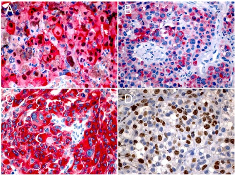
Study design: Twenty two cases of primary oral melanoma were analyzed by description of their histopathological features and immunohistochemical study using the antibodies S-100, HMB-45, Melan-A and Ki-67.
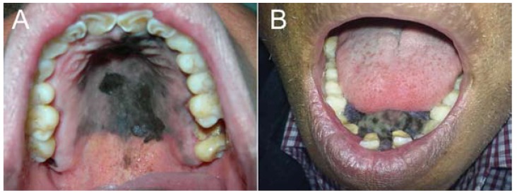
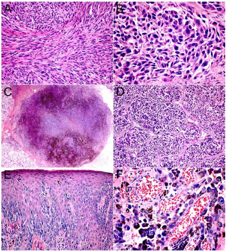
Results: The mean age was 58 years and 14 cases were female. The main affected sites were the hard palate, followed by the upper gingiva. Microscopically, 15 cases presented level III of invasion, 2 cases were amelanotic and 13 showed a mixed epithelioid and plasmacytoid or spindle cells composition. Some cases showed necrosis, perivascular and perineural invasion. S-100 and HMB-45 were positive in all cases, but 3 cases were negative for Melan-A. The proliferative index with Ki-67 was high, with labeling index ranging from 15.51% to 63% of positive cells.
Conclusion: S-100 and HMB-45 are more frequently expressed than Melan-A in primary oral melanomas and these markers are helpful to confirm the diagnosis.
Figures
Similar articles
-
[Perivascular epithelioid cell tumor of urinary system: a clinicopathologic analysis of 21 cases].Zhonghua Bing Li Xue Za Zhi. 2012 Jul;41(7):443-7. doi: 10.3760/cma.j.issn.0529-5807.2012.07.003. Zhonghua Bing Li Xue Za Zhi. 2012. PMID: 22932453 Chinese.
-
Immunohistochemical diagnosis of malignant melanoma of the conjunctiva and uvea: comparison of the novel antibody against melan-A with S100 protein and HMB-45.Melanoma Res. 2000 Aug;10(4):350-4. doi: 10.1097/00008390-200008000-00006. Melanoma Res. 2000. PMID: 10985669
-
Immunohistochemical studies of conjunctival nevi and melanomas.Arch Ophthalmol. 2010 Feb;128(2):174-83. doi: 10.1001/archophthalmol.2009.394. Arch Ophthalmol. 2010. PMID: 20142539
-
Immunohistochemical characteristics of melanoma.J Cutan Pathol. 2008 May;35(5):433-44. doi: 10.1111/j.1600-0560.2007.00891.x. J Cutan Pathol. 2008. PMID: 18399807 Review.
-
Primary mucosal malignant melanoma of the cervix: case report and review of the literature.Tumori. 2015 Sep 9;101(5):e147-50. doi: 10.5301/tj.5000304. Tumori. 2015. PMID: 25983102 Review.
Cited by
-
Oral pigmented lesions: a retrospective analysis from Brazil.Med Oral Patol Oral Cir Bucal. 2021 May 1;26(3):e284-e291. doi: 10.4317/medoral.24168. Med Oral Patol Oral Cir Bucal. 2021. PMID: 32856618 Free PMC article.
-
Amelanotic Melanoma Presenting with Plasmacytoid Morphology and BRAF V600 Mutation.Rare Tumors. 2015 Jun 25;7(2):5698. doi: 10.4081/rt.2015.5698. eCollection 2015 May 5. Rare Tumors. 2015. PMID: 26266008 Free PMC article.
-
Certainty of S100 from Physiology to Pathology.J Clin Diagn Res. 2016 Jun;10(6):ZE10-5. doi: 10.7860/JCDR/2016/17949.8022. Epub 2016 Jun 1. J Clin Diagn Res. 2016. PMID: 27504432 Free PMC article. Review.
-
Oral Malignant Melanoma Initially Misdiagnosed as a Racial Pigmentation: A Case Report.Dermatopathology (Basel). 2016 Feb 26;3(1):1-7. doi: 10.1159/000444049. eCollection 2016 Jan-Mar. Dermatopathology (Basel). 2016. PMID: 27195264 Free PMC article.
-
Clinicopathological and prognostic value of Ki-67 expression in oral malignant melanoma: A systematic review and meta-analysis.J Dent Res Dent Clin Dent Prospects. 2022 Summer;16(3):140-146. doi: 10.34172/joddd.2022.024. Epub 2022 Nov 15. J Dent Res Dent Clin Dent Prospects. 2022. PMID: 36704188 Free PMC article. Review.
References
-
- Femiano F, Lanza A, Buonaiuto C, Gombos F, Di Spirito F, Cirillo N. Oral malignant melanoma: a review of the literature. J Oral Pathol Med. 2008;37:383–8. - PubMed
-
- Hashemi Pour MS. Malignant melanoma of the oral cavity: a review of literature. Indian J Dent Res. 2008;19:47–51. - PubMed
-
- Gu GM, Epstein JB, Morton TH. Intraoral melanoma: long-term follow-up and implication for dental clinicians. A case report and literature review. Oral Surg Oral Med Oral Pathol Oral Radiol Endod. 2003;96:404–13. - PubMed
-
- Hicks MJ, Flaitz CM. Oral mucosal melanoma: epidemiology and pathobiology. Oral Oncol. 2000;36:152–69. - PubMed
-
- Meleti M, Leemans CR, Mooi WJ, Vescovi P, van der Waal I. Oral malignant melanoma: a review of the literature. Oral Oncol. 2007;43:116–21. - PubMed
MeSH terms
Substances
LinkOut - more resources
Full Text Sources
Medical
