. 2011 Dec 20;108(51):20562-7.
doi: 10.1073/pnas.1115124108. Epub 2011 Dec 5.

Computational design of a symmetric homodimer using β-strand assembly

Affiliations

Computational design of a symmetric homodimer using β-strand assembly

P Benjamin Stranges et al. Proc Natl Acad Sci U S A. .

Abstract

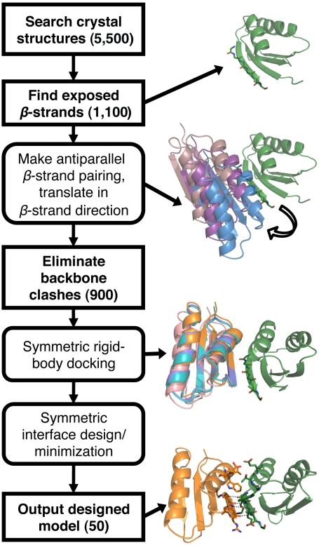
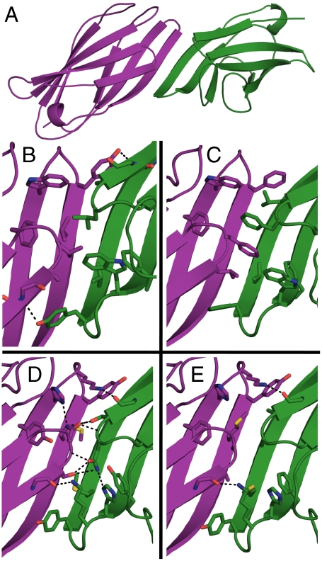
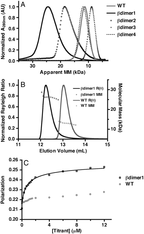
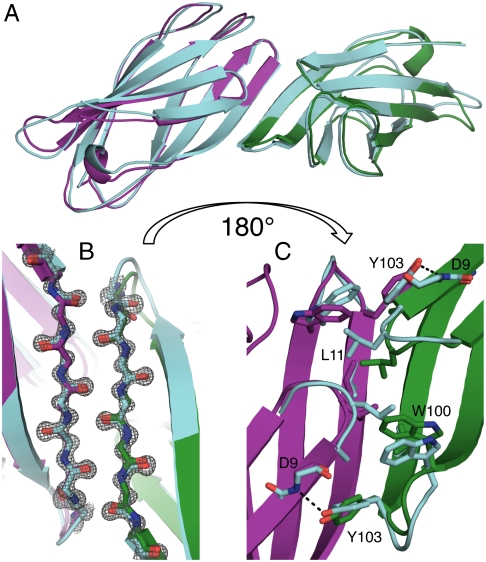
Computational design of novel protein-protein interfaces is a test of our understanding of protein interactions and has the potential to allow modification of cellular physiology. Methods for designing high-affinity interactions that adopt a predetermined binding mode have proved elusive, suggesting the need for new strategies that simplify the design process. A solvent-exposed backbone on a β-strand is thought of as "sticky" and β-strand pairing stabilizes many naturally occurring protein complexes. Here, we computationally redesign a monomeric protein to form a symmetric homodimer by pairing exposed β-strands to form an intermolecular β-sheet. A crystal structure of the designed complex closely matches the computational model (rmsd = 1.0 Å). This work demonstrates that β-strand pairing can be used to computationally design new interactions with high accuracy.


Conflict of interest statement

The authors declare no conflict of interest.

Figures

Fig. 1.
Fig. 1.
Search and design protocol for a symmetric β-strand mediated homodimer. Method used to search for, then design, scaffold proteins to create a symmetric homodimer (see full details in Materials and Methods). Numbers in parentheses represent the total number of unique input structures used in each step. Individual steps are illustrated by the structures generated during each step using the protein Atx1 (Protein Data Bank ID 1CC8).
Fig. 2.
Fig. 2.
Computational designs used in experiments. (A) Overall topology of computational designs. The γ-adaptin appendage domain (Protein Data Bank ID 2A7B) is used as the scaffold for the designed interface. Coloring (purple and green) highlights the symmetric chains in the model. The solvent-excluded side of the interface is shown in detail for βdimer1 (B), βdimer2 (C), βdimer3 (D), and βdimer4 (E). Selected side chains are shown in sticks. Black dashed lines represent hydrogen bonds at the interface; the six main-chain hydrogen bonds are not shown.
Fig. 3.
Fig. 3.
Experimental determination of molecular mass in solution. (A) Size-exclusion chromatography (Superdex 75) of the designs and wild-type protein. Absorbance has been normalized based on maximum value, the apparent molecular mass (MM) is based on a standard curve obtained from globular proteins. (B) Size-exclusion chromatography (Superdex 75) followed by multiangle light scattering of wild type (gray) and βdimer1 (black). Rayleigh ratio [R(θ)] (solid lines) has been normalized based on maximum value; MM (open circles) is calculated from light scattering and refractive index. The average molecular mass is 26 kDa for βdimer1, 14 kDa for the wild type. (C) Measurement of dimer dissociation constant of βdimer1 using a fluorescence polarization assay. Bodipy-labeled βdimer1 was titrated with unlabeled βdimer1 (black) and wild-type protein (gray), and the change in polarization was fit to a homodimerization model (see SI Materials and Methods). The calculated homodimer dissociation constant for βdimer1 is 1.0 ± 0.1 μM (SEM).
Fig. 4.
Fig. 4.
Comparison of βdimer1 computational model to crystal structure. (A) Overlay of the βdimer1 computational model (green and purple) and crystal structure (cyan). The backbone atom rmsd for the entire structure is 1.0 Å. (B) Backbone–backbone interactions between the interface-forming β-strands viewed from the solvent-accessible side of the intermolecular β-sheet. The 2Fo - Fc electron density (gray) is contoured to 2σ. (C) Detailed view of designed side chains forming interactions on the solvent-excluded side of interacting β-strands. A black dashed line represents the interface-spanning hydrogen-bond between D9 and Y103.

Similar articles

Cited by

References

    1. Karanicolas J, Kuhlman B. Computational design of affinity and specificity at protein–protein interfaces. Curr Opin Struct Biol. 2009;19:458–463. - PMC - PubMed
    1. Mandell DJ, Kortemme T. Computer-aided design of functional protein interactions. Nat Chem Biol. 2009;5:797–807. - PubMed
    1. Havranek JJ, Harbury PB. Automated design of specificity in molecular recognition. Nat Struct Biol. 2003;10:45–52. - PubMed
    1. Grigoryan G, et al. Computational design of virus-like protein assemblies on carbon nanotube surfaces. Science. 2011;332:1071–1076. - PMC - PubMed
    1. Grigoryan G, Reinke AW, Keating AE. Design of protein-interaction specificity gives selective bZIP-binding peptides. Nature. 2009;458:859–864. - PMC - PubMed

Publication types

Associated data

LinkOut - more resources