. 2011 Dec 20;108(51):20491-6.
doi: 10.1073/pnas.1111014108. Epub 2011 Dec 5.

Multiple molecular architectures of the eye lens chaperone αB-crystallin elucidated by a triple hybrid approach

Affiliations

Multiple molecular architectures of the eye lens chaperone αB-crystallin elucidated by a triple hybrid approach

Nathalie Braun et al. Proc Natl Acad Sci U S A. .

Abstract

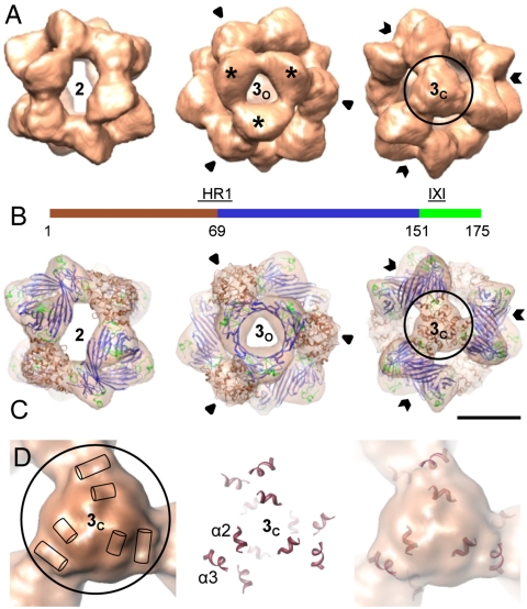
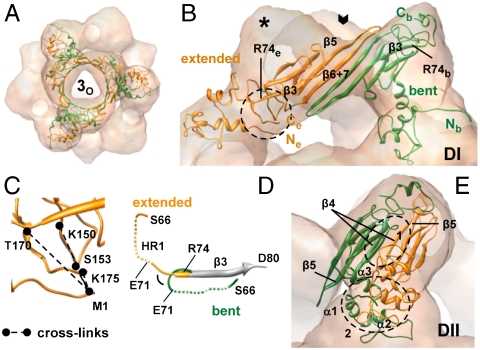
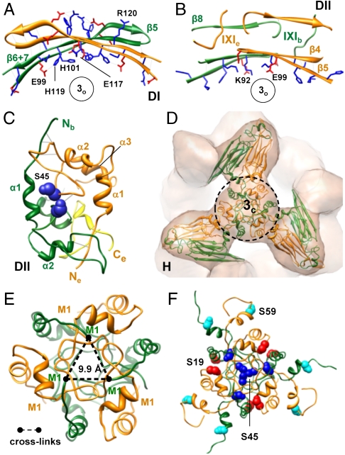
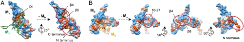
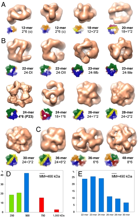
The molecular chaperone αB-crystallin, the major player in maintaining the transparency of the eye lens, prevents stress-damaged and aging lens proteins from aggregation. In nonlenticular cells, it is involved in various neurological diseases, diabetes, and cancer. Given its structural plasticity and dynamics, structure analysis of αB-crystallin presented hitherto a formidable challenge. Here we present a pseudoatomic model of a 24-meric αB-crystallin assembly obtained by a triple hybrid approach combining data from cryoelectron microscopy, NMR spectroscopy, and structural modeling. The model, confirmed by cross-linking and mass spectrometry, shows that the subunits interact within the oligomer in different, defined conformations. We further present the molecular architectures of additional well-defined αB-crystallin assemblies with larger or smaller numbers of subunits, provide the mechanism how "heterogeneity" is achieved by a small set of defined structural variations, and analyze the factors modulating the oligomer equilibrium of αB-crystallin and thus its chaperone activity.


Conflict of interest statement

The authors declare no conflict of interest.

Figures

Fig. 1.
Fig. 1.
Three-dimensional model of the αB-crystallin 24-mer. (A) Surface representations of the cryo-EM density map viewed along a two- (Left) and a threefold symmetry axis intercepting the area harboring a “window” (3o, open arrows) (Center), and mass accumulation (3c, closed arrows) (Right). Mass-rich domes surrounding 3o are highlighted by stars. The isosurface threshold was set to enclose a molecular mass of 485 kDa. (B) Domain organization of human αB-crystallin: N-terminal segment (residues 1–68) (brown), ACD (residues 69–150) (blue), C-terminal region (residues 151–175) (lime green). The heterogeneous region 1 (HR1) and the IXI motif are indicated. (C) Views of the oligomer with the docked hybrid model of αB-crystallin 24-mer (ribbon representation) superimposed. Ribbon color coding is the same as in B. (Scale bar, 5 nm.) (D) Close-up view of the density map at the area 3c (Left). The positions of the rod-like densities are schematically indicated by cylinders. Surface near helices in the pseudoatomic model (Center) superposed on the cryo-EM density map (Right).
Fig. 2.
Fig. 2.
The αB-crystallin adopts in the 24-mer two different conformations. (A) The αB-crystallin hexamer (ribbon diagram) viewed along a threefold symmetry axis. Conformationally different monomers extended (Me) and bent (Mb) are shown in orange and green, respectively. (B) Spatial arrangement of Me and Mb in type I dimer (DI). Open arrow indicates the position of a threefold axis (3o). The residue R74 is labeled on both conformers. The area highlighted by a circle encloses the N and C termini of Me. (C) Enlarged view of the encircled area in B with the intramolecular cross-links between M1 and C-terminal residues of Me. (D) Close-up view of the region S66-D80. The β3 strands (residues 74–79) of Me and Mb are aligned and partly shown in gray. Dashed lines: HR1 regions (E67–L70 corresponding to the β2 strand in other sHsps) of Me and Mb. Note the different environments for HR1 in both conformers. (E) Spatial arrangement of Me and Mb in type II dimer (DII). Close-up views of DII interface patches 1 and 2 highlighted by circles are shown in Fig. 3.
Fig. 3.
Fig. 3.
Subunit interfaces in the αB-crystallin 24-mer. (A) DI interface viewed along a threefold axis (for the spatial arrangement and localization of the type I dimer see Fig. 2B). Positively and negatively charged residues of the β5 and β6 + 7 strands are shown in blue and red, respectively. (B) DII interface patch formed by the ACDs of both conformers (area 1 in Fig. 2E). Charged residues of the β4 and β5 strands and segments of both protomers containing the IXI motifs are shown. (C) DII interface patch encompassing the N- and C-terminal domains of both conformers (area 2 in Fig. 2E). S45 residues of both conformers are shown as spheres in dark blue. The C terminus of the extended monomer is colored yellow. (D) Hexamer assembly site at the area 3c. For close-up views of the encircled area see E and F. (E) Intermolecular cross-links between the N termini of bent monomers at the hexamer assembly site. (F) Location of the N-terminal serine residues within the hexamer assembly area. The serine residues 19, 45, and 59 are shown as spheres and are colored in red, dark blue, and cyan, respectively.
Fig. 4.
Fig. 4.
Hydrophobic patches of αB-crystallin buried at subunit interfaces. (A) Hydrophobic patches of an extended monomer (Me) (hydrophobicity surface) buried by the overlying bent monomer (Mb) (ribbon diagram, green) at the DII interface of the 24-mer. (B) Hydrophobic patches of a bent monomer (Mb) (hydrophobicity surface) exposed upon removal of the overlying extended monomer (Me) (ribbon diagram, orange). Color coding: hydrophilic, blue; neutral, white; hydrophobic, orange.
Fig. 5.
Fig. 5.
Three-dimensional reconstructions of αB-crystallin oligomers and their distribution. Oligomers found in the “small” dataset (A), in the “24-mer” dataset (B), and in the “large” dataset (C). (Insets) Oligomer models showing the boundaries of hexameric and dimeric building blocks. The missing volume of the monomer in the 23-mers in B is highlighted in mesh representation. (D) Distribution of the oligomer masses derived from particle size and form distributions obtained upon 4D projection matching cycles applied to datasets small, 24-mer, and large. Particles with molecular masses (MM) corresponding to 12-, 18-, and 20-mers are included in green bars, to the 24-mer in the blue bar, to 36- and 48-mers in red bars. (E) Distribution of oligomers within the supposed 24-mer population (blue bar in D) determined by 4D projection matching cycles using all 22- to 30-mer pseudoatomic models.

Similar articles

Cited by

References

    1. Bloemendal H, et al. Ageing and vision: Structure, stability and function of lens crystallins. Prog Biophys Mol Biol. 2004;86:407–485. - PubMed
    1. Delaye M, Tardieu A. Short-range order of crystallin proteins accounts for eye lens transparency. Nature. 1983;302:415–417. - PubMed
    1. Horwitz J. α-crystallin can function as a molecular chaperone. Proc Natl Acad Sci USA. 1992;89:10449–10453. - PMC - PubMed
    1. Bloemendal H. The vertebrate eye lens. A useful system for the study of fundamental biological processes on a molecular level. Science. 1977;197:127–138. - PubMed
    1. de Jong WW, Caspers GJ, Leunissen JAM. Genealogy of the α-crystallin-small heat-shock protein superfamily. Int J Biol Macromol. 1998;22:151–162. - PubMed

Publication types

MeSH terms

Associated data

LinkOut - more resources