. 2011 Dec 20;108(51):20503-8.
doi: 10.1073/pnas.1112831108. Epub 2011 Dec 5.

Sterol-induced degradation of HMG CoA reductase depends on interplay of two Insigs and two ubiquitin ligases, gp78 and Trc8

Affiliations

Sterol-induced degradation of HMG CoA reductase depends on interplay of two Insigs and two ubiquitin ligases, gp78 and Trc8

Youngah Jo et al. Proc Natl Acad Sci U S A. .

Abstract

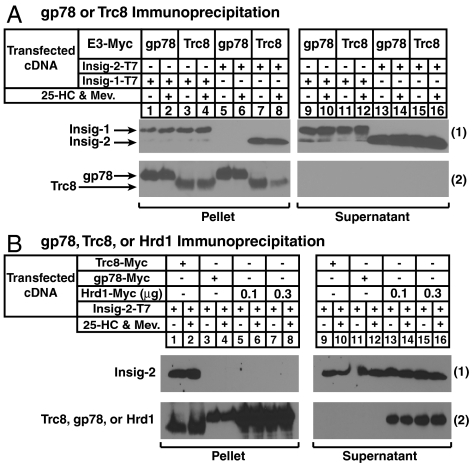
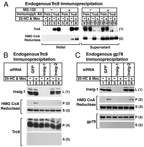
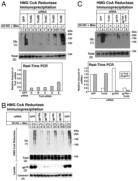
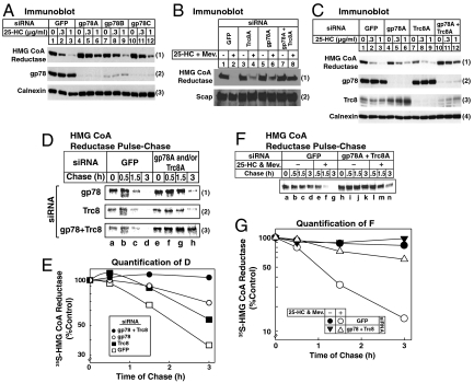
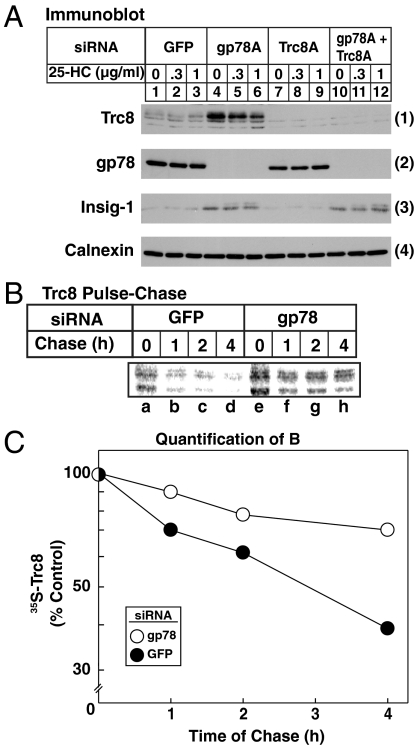
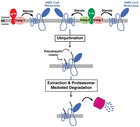
Accumulation of sterols in membranes of the endoplasmic reticulum (ER) leads to the accelerated ubiquitination and proteasomal degradation of 3-hydroxy-3-methylglutaryl coenzyme A reductase, a rate-limiting enzyme in synthesis of cholesterol and nonsterol isoprenoids. This degradation results from sterol-induced binding of reductase to the Insig-1 or Insig-2 proteins of ER membranes. We previously reported that in immortalized human fibroblasts (SV-589 cells) Insig-1, but not Insig-2, recruits gp78, a membrane-bound RING-finger ubiquitin ligase. We now report that both Insig-1 and Insig-2 bind another membrane-bound RING-finger ubiquitin ligase called Trc8. Knockdown of either gp78 or Trc8 in SV-589 cells through RNA interference (RNAi) inhibited sterol-induced ubiquitination of reductase and inhibited sterol-induced degradation by 50-60%. The combined knockdown of gp78 and Trc8 produced a more complete inhibition of degradation (> 90%). Knockdown of gp78 led to a three to fourfold increase in levels of Trc8 and Insig-1 proteins, which opposed the inhibitory action of gp78. In contrast, knockdown of Trc8 had no effect on gp78 or Insig-1. The current results suggest that sterol-induced ubiquitination and proteasomal degradation of reductase is dictated by the complex interplay of at least four proteins: Insig-1, Insig-2, gp78, and Trc8. Variations in the concentrations of any one of these proteins may account for differences in cell- and/or tissue-specific regulation of reductase degradation.


Conflict of interest statement

The authors declare no conflict of interest.

Figures

Fig. 1.
Fig. 1.
Association of Insig-1 and Insig-2 with membrane-bound ubiquitin ligases. (A) CHO-7 cells were set up on day 0 at 5 × 105 cells per 60-mm dish in medium A containing 5% LPDS. On day 1, cells were transfected in 5% LPDS with 50 ng/dish of pCMV-Insig-1-T7, 500 ng/dish of pCMV-Insig-2-T7, 3 ng/dish of pCMV-gp78-Myc, and 30 ng/dish of pCMV-Trc8-Myc as indicated and described in Materials and Methods. The total amount of DNA/dish was adjusted to 3 μg by the addition of empty pcDNA3 vector. Following incubation for 6 h at 37 °C, cells were depleted of sterols by direct addition of medium A containing 5% LPDS, 10 μM sodium compactin, and 50 μM sodium mevalonate (final concentration). After 16 h at 37 °C, cells were refed identical medium containing 10 μM MG-132 in the absence or presence of 1 μg/mL 25-HC plus 10 mM mevalonate and incubated for an additional 5 h. Cells were subsequently harvested for preparation of detergent lysates that were immunoprecipitated with anti-Myc-coupled agarose beads. Immunoprecipitation pellet and supernatant fractions were subjected to SDS-PAGE and immunoblot analysis with anti-T7 IgG (against Insig-1 and Insig-2) or IgG-9E10 (against gp78 and Trc8). (B) CHO-7 cells were set up on day 0, transfected with pCMV-Insig-2-T7 (150 ng), pCMV-Trc8-Myc (100 ng), pCMV-gp78-Myc (10 ng), and pCMV-Hrd1-Myc (0.3 and 1 μg) as indicated, depleted of sterols on day 1, and treated with 25-HC plus mevalonate on day 2 as described in (A). Following treatments, cells were harvested for immunoprecipitation with anti-Myc-coupled agarose beads. Resulting pellet and supernatant fractions were immunoblotted with anti-T7 IgG (against Insig-2) or IgG-9E10 (against Trc8, gp78, and Hrd1).
Fig. 2.
Fig. 2.
Sterol-induced, Insig-mediated binding of Trc8 to HMG CoA reductase. (A) SV-589 cells were set up on day 0 at 2 × 105 cells per 100-mm dish in medium B supplemented with 10% FCS. On day 3, cells were depleted of sterols through incubation for 16 h at 37 °C in medium B containing 10% LPDS, 50 μM compactin, and 50 μM mevalonate. The cells were then switched to identical medium containing 10 μM MG-132 for 2 h, after which they received 1 μg/mL 25-HC plus 10 mM mevalonate as indicated and incubated for an additional 2 h at 37 °C. Cells were then harvested and immunoprecipitated with IgG-556 (against Trc8); resulting pellet and supernatant fractions were subjected to immunoblot analysis with IgG-3D10 (against Trc8) and IgG-A9 (against reductase). (B and C) SV-589 cells were set up on day 0 as described in (A). On days 1 and 2, the cells were transfected in medium B containing 10% FCS with the indicated siRNA as described in Materials and Methods. After the second transfection on day 2, cells were depleted of sterols as described in (A) and then switched to identical medium containing 10 μM MG-132 in the absence or presence of 1 μg/mL 25-HC plus 10 mM mevalonate. Following incubation for 2 h at 37 °C, cells were harvested and lysates subjected to immunoprecipitation with either IgG-556 [against Trc8, (B)] or IgG-740F [against gp78, (C)]. Resulting pellet (P) and supernatant (S) fractions as well as total lysates (L) were subjected to immunoblot analysis with IgG-3D10 (against Trc8), IgG-A9 (against reductase), and IgG-740F, and IgG-17H1 (against Insig-1). The two bands for Insig-1 result from alternative use of initiating methionines (21). The larger band results from translation initiation at residue 1, whereas the smaller band results from initiation at residue 37.
Fig. 3.
Fig. 3.
Inhibition of sterol-induced ubiquitination of HMG CoA reductase by RNA interference-mediated knockdown of Trc8 and gp78. SV-589 cells were set up on day 0, transfected with the indicated siRNAs on days 1 and 2, and depleted of sterols as described in Fig. 2A. Sterol-depleted cells were subsequently incubated for 30 min in medium B containing 10% LPDS, 50 μM compactin, and 10 μM MG-132 in the absence or presence of 1 μg/mL 25-HC plus 10 mM mevalonate as indicated. Cells were harvested and immunoprecipitated with polyclonal anti-reductase, followed by immunoblot analysis with IgG-A9 (against reductase) and IgG-P4D1 (against ubiquitin). Total RNA from 25-HC plus mevalonate-treated cells in (A) and (C) was subjected to first-strand cDNA synthesis and real-time PCR. Each value for cells transfected with the indicated siRNA represents the amount of the indicated mRNA relative to that in control cells transfected with the GFP siRNA.
Fig. 4.
Fig. 4.
Trc8 and gp78 are required for sterol-accelerated degradation of HMG CoA reductase as revealed by RNA interference. SV-589 cells were set up for experiments on day 0, transfected with the indicated siRNAs on days 1 and 2 (A, C, D, and F) or on days 1 and 3 (B), and depleted of sterols as described in Fig. 2A. (AC) Sterol-depleted cells were incubated for 2 h in medium B supplemented with 10% LPDS and 50 μM compactin in the absence or presence of the indicated concentration of 25-HC in (A and C) or 0.5 μg/mL 25-HC plus 10 mM mevalonate in (B). Following incubation for 2–3 h at 37 °C, cells were harvested for subcellular fractionation. Aliquots of the membrane fractions (normalized for equal protein loaded/lane) were subjected to SDS-PAGE and immunoblot analysis with IgG-A9 (against reductase), IgG-R139 (against Scap), IgG-740F (against gp78), IgG-556 (against Trc8), and anti-calnexin IgG. (D and F) Sterol-depleted cells were preincubated with methionine/cysteine-free medium B containing 10% LPDS and 50 μM compactin for 1 h at 37 °C. After pulse-labeling for 30 min at 37 °C in identical medium containing 130 μCi/mL of [35S] methionine, cells were chased in medium B supplemented with 10% LPDS, 50 μM compactin, 0.5 mM unlabeled methionine, and 1 mM cysteine in the absence or presence of 1 μg/mL 25-HC plus 10 mM mevalonate as indicated. Following the indicated time of chase, cells were harvested and subjected to anti-reductase immunoprecipitation, followed by SDS-PAGE and transfer of proteins to a nitrocellulose filter. The filter was exposed to an imaging plate at room temperature, scanned in a Storm 820 PhosphorImager, and the image was photographed. (E and G) [35S] Radioactivity in the scanned gels from Fig. 4 D and F corresponding to reductase was quantified by densitometry. The intensity of reductase during the pulse [lanes a and e in (D); lanes a and h in (F)] was arbitrarily set at 100%.
Fig. 5.
Fig. 5.
Stabilization of Trc8 by RNA interference-mediated knockdown of gp78. SV-589 cells were set up for experiments on day 0, transfected with the indicated siRNAs on days 1 and 2 as described in Fig. 3. (A) Following sterol depletion, cells were incubated in medium B supplemented with 10% LPDS, 50 μM compactin, and 50 μM cycloheximide. After 1 h at 37 °C, cells received the indicated concentration of 25-HC and incubated an additional 2 h. Cells were subsequently harvested for subcellular fractionation; resulting membrane fractions were subjected to immunoblot analysis with IgG-556 (against Trc8), IgG-740F (against gp78), IgG-17H1 (against Insig-1), and anti-calnexin IgG. Results are representative of at least two independent experiments. (B) Cells were pulse-labeled for 30 min in medium B containing 10% FCS and 130 μCi/mL [35S] methionine and subsequently chased in medium B supplemented with 10% FCS, 0.5 mM unlabeled methionine, and 1 mM cysteine. Following the indicated time of chase, cells were harvested, subjected to anti-Trc8 immunoprecipitation, and analyzed as described in Fig. 4. (C) [35S] Radioactivity in the scanned gel from Fig. 5B corresponding to the two bands of Trc8 was quantified by densitometry. The nature of these two bands is currently unknown. The intensity of reductase during the pulse (Fig. 5B, lanes a and e) was arbitrarily set at 100%.
Fig. 6.
Fig. 6.
Model for sterol-accelerated ubiquitination and degradation of HMG CoA reductase mediated by Trc8 and gp78. In sterol-treated cells, Insig-1 bridges gp78 to reductase and either Insig-1 or Insig-2 bridges Trc8 to reductase for ubiquitination. The resultant gp78- and Trc8-mediated ubiquitination marks reductase for extraction from ER membranes and subsequent delivery to proteasomes for degradation.

Similar articles

Cited by

References

    1. DeBose-Boyd RA. Feedback regulation of cholesterol synthesis: sterol-accelerated ubiquitination and degradation of HMG CoA reductase. Cell Res. 2008;18:609–621. - PMC - PubMed
    1. Brown MS, Goldstein JL. Multivalent feedback regulation of HMG CoA reductase, a control mechanism coordinating isoprenoid synthesis and cell growth. J Lipid Res. 1980;21:505–517. - PubMed
    1. Sever N, Yang T, Brown MS, Goldstein JL, DeBose-Boyd RA. Accelerated degradation of HMG CoA reductase mediated by binding of insig-1 to its sterol-sensing domain. Mol Cell. 2003;11:25–33. - PubMed
    1. Sever N, et al. Insig-dependent ubiquitination and degradation of mammalian 3-hydroxy-3-methylglutaryl-CoA reductase stimulated by sterols and geranylgeraniol. J Biol Chem. 2003;278:52479–52490. - PubMed
    1. Liscum L, et al. Domain structure of 3-hydroxy-3-methylglutaryl coenzyme A reductase, a glycoprotein of the endoplasmic reticulum. J Biol Chem. 1985;260:522–530. - PubMed

Publication types

MeSH terms

LinkOut - more resources