

Multimodal image coregistration and inducible selective cell ablation to evaluate imaging ligands
- PMID: 22143775
- PMCID: PMC3251115
- DOI: 10.1073/pnas.1109480108
Multimodal image coregistration and inducible selective cell ablation to evaluate imaging ligands
Abstract
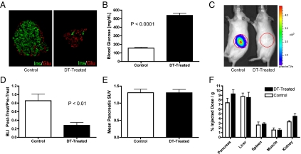
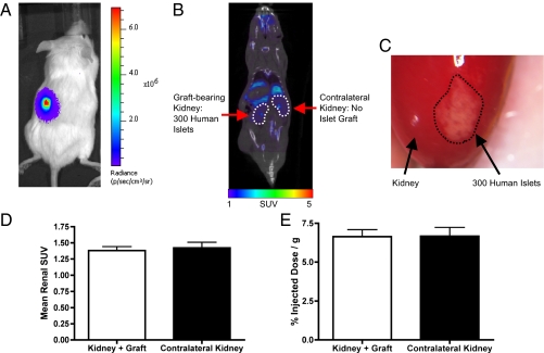
We combined multimodal imaging (bioluminescence, X-ray computed tomography, and PET), tomographic reconstruction of bioluminescent sources, and two unique, complementary models to evaluate three previously synthesized PET radiotracers thought to target pancreatic beta cells. The three radiotracers {[(18)F]fluoropropyl-(+)-dihydrotetrabenazine ([(18)F]FP-DTBZ), [(18)F](+)-2-oxiranyl-3-isobutyl-9-(3-fluoropropoxy)-10-methoxy-2,3,4,6,7,11b-hexahydro-1H-pyrido[2,1-a]isoquinoline ((18)F-AV-266), and (2S,3R,11bR)-9-(3-fluoropropoxy)-2-(hydroxymethyl)-3-isobutyl-10-methoxy-2,3,4,6,7,11b-hexahydro-1H-pyrido[2,1-a]isoquinolin-2-ol ((18)F-AV-300)} bind vesicular monoamine transporter 2. Tomographic reconstruction of the bioluminescent signal in mice expressing luciferase only in pancreatic beta cells was used to delineate the pancreas and was coregistered with PET and X-ray computed tomography images. This strategy enabled unambiguous identification of the pancreas on PET images, permitting accurate quantification of the pancreatic PET signal. We show here that, after conditional, specific, and rapid mouse beta-cell ablation, beta-cell loss was detected by bioluminescence imaging but not by PET imaging, given that the pancreatic signal provided by three PET radiotracers was not altered. To determine whether these ligands bound human beta cells in vivo, we imaged mice transplanted with luciferase-expressing human islets. The human islets were imaged by bioluminescence but not with the PET ligands, indicating that these vesicular monoamine transporter 2-directed ligands did not specifically bind beta cells. These data demonstrate the utility of coregistered multimodal imaging as a platform for evaluation and validation of candidate ligands for imaging islets.
Conflict of interest statement
Conflict of interest statement: F.H., D.S., and H.F.K. either are employed at Avid Radiopharmaceuticals or have a financial interest in the company. A.C.P. has received an investigator-initiated research grant from Eli Lilly and Company.
Figures

References
-
- Calne DB, et al. Positron emission tomography after MPTP: Observa-tions relating to the cause of Parkinson's disease. Nature. 1985;317:246–248. - PubMed
-
- Som P, et al. A fluorinated glucose analog, 2-fluoro-2-deoxy-d-glucose (F-18): Nontoxic tracer for rapid tumor detection. J Nucl Med. 1980;21:670–675. - PubMed
-
- Robertson RP. Estimation of β-cell mass by metabolic tests: Necessary, but how sufficient? Diabetes. 2007;56:2420–2424. - PubMed
-
- Gepts W. Pathologic anatomy of the pancreas in juvenile diabetes mellitus. Diabetes. 1965;14:619–633. - PubMed
Publication types
MeSH terms
Substances
Grants and funding
- DK072473/DK/NIDDK NIH HHS/United States
- DK69603/DK/NIDDK NIH HHS/United States
- R21 DK068854/DK/NIDDK NIH HHS/United States
- U24CA126588/CA/NCI NIH HHS/United States
- DK089572/DK/NIDDK NIH HHS/United States
- U01 DK089572/DK/NIDDK NIH HHS/United States
- R33 DK066636/DK/NIDDK NIH HHS/United States
- R01 DK069603/DK/NIDDK NIH HHS/United States
- R01 DK068764/DK/NIDDK NIH HHS/United States
- DK68854/DK/NIDDK NIH HHS/United States
- DK20593/DK/NIDDK NIH HHS/United States
- U24 DK059637/DK/NIDDK NIH HHS/United States
- P60 DK020593/DK/NIDDK NIH HHS/United States
- T32 EB001628/EB/NIBIB NIH HHS/United States
- U24 CA126588/CA/NCI NIH HHS/United States
- U01 DK072473/DK/NIDDK NIH HHS/United States
- DK68764/DK/NIDDK NIH HHS/United States
- DK66636/DK/NIDDK NIH HHS/United States
- I01 BX000666/BX/BLRD VA/United States
- DK59637/DK/NIDDK NIH HHS/United States
- P30 DK020593/DK/NIDDK NIH HHS/United States
- T32EB001628/EB/NIBIB NIH HHS/United States
- R21 DK066636/DK/NIDDK NIH HHS/United States
LinkOut - more resources
Full Text Sources
Other Literature Sources
Medical
