. 2011 Dec 20;108(51):20684-9.
doi: 10.1073/pnas.1116848108. Epub 2011 Dec 5.

Sterile inflammation of endothelial cell-derived apoptotic bodies is mediated by interleukin-1α

Affiliations

Sterile inflammation of endothelial cell-derived apoptotic bodies is mediated by interleukin-1α

Yaël Berda-Haddad et al. Proc Natl Acad Sci U S A. .

Abstract

Sterile inflammation resulting from cell death is due to the release of cell contents normally inactive and sequestered within the cell; fragments of cell membranes from dying cells also contribute to sterile inflammation. Endothelial cells undergoing stress-induced apoptosis release membrane microparticles, which become vehicles for proinflammatory signals. Here, we show that stress-activated endothelial cells release two distinct populations of particles: One population consists of membrane microparticles (<1 μm, annexin V positive without DNA and no histones) and another larger (1-3 μm) apoptotic body-like particles containing nuclear fragments and histones, representing apoptotic bodies. Contrary to present concepts, endothelial microparticles do not contain IL-1α and do not induce neutrophilic chemokines in vitro. In contrast, the large apoptotic bodies contain the full-length IL-1α precursor and the processed mature form. In vitro, these apoptotic bodies induce monocyte chemotactic protein-1 and IL-8 chemokine secretion in an IL-1α-dependent but IL-1β-independent fashion. Injection of these apoptotic bodies into the peritoneal cavity of mice induces elevated serum neutrophil-inducing chemokines, which was prevented by cotreatment with the IL-1 receptor antagonist. Consistently, injection of these large apoptotic bodies into the peritoneal cavity induced a neutrophilic infiltration that was prevented by IL-1 blockade. Although apoptosis is ordinarily considered noninflammatory, these data demonstrate that nonphagocytosed endothelial apoptotic bodies are inflammatory, providing a vehicle for IL-1α and, therefore, constitute a unique mechanism for sterile inflammation.


Conflict of interest statement

The authors declare no conflict of interest.

Figures

Fig. 1.
Fig. 1.
Effect of serum starvation and stimulation with TNFα on apoptosis in HUVEC. (A) HUVEC cellular and nuclear morphology observed by light (ac) or fluorescent microscopy after DAPI labeling (a’–c’). HUVEC were cultured in normal condition (N) or in starved conditions (STV) for 16 h, then stimulated with TNFα for 24 h (STV+TNFα); white arrows show cell nucleus fragmentation. (B) Percentage of AnnV-FITC and PI double-labeled HUVEC in each condition (*P < 0.05, n = 4). (C) Western blot of caspase-3 cleavage products in HUVEC under the conditions in A. The 17- to 19-kDa band representing the larger form of cleaved caspase 3 is observed in STV and STV+TNFα. Shown is one representative blot of three experiments. (D) Presence of cleaved caspase 3 using fluorescence microscopy in starved TNFα stimulated HUVEC. HUVEC were labeled with anti-cleaved caspase-3 mAb, and nuclei were counterstained with DAPI. One representative among three experiments.
Fig. 2.
Fig. 2.
Characterization of fractions of particles in supernatants from stress-induced apoptosis in HUVEC. Supernatants of starved HUVEC (10 × 106 cells) stimulated with TNFα were centrifuged at 4,500 x g and 75,000 x g. (A) AnnV-FITC–positive particles from 4,500 x g and 75,000 x g pellets have different size distribution as estimated by forward scatter in flow cytometry. The y axis represents forward scatter. The x axis represents structure scatter. One representative among three different experiments. (B) Double staining using AnnV-FITC and DAPI separate the different particulate population from 4,500 x g and 75,000 x g pellets. Shown is one representative among three different experiments. (C) Western blotting of 4,500 x g and 75,000 x g pellets were analyzed by immunoblotting for the detection of histones using anti-H3 and anti–p-H2B Ab.
Fig. 3.
Fig. 3.
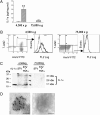
Detection of IL-1α in AptB but not in MP. (A) A concentration of 30.5 ± 5.3 pg of IL-1α /mg of protein (50 μg were used in all experiments) was measured by using specific ELISA in 4,500 x g pellet from TNFα-stimulated starved HUVEC (hatched bars) versus 1.2 ± 1 pg/mg in unstimulated starved HUVEC (open bars) (**P < 0.006, n = 3). In the 75,000 x g pellet, IL-1α was weakly detected in starved TNFα HUVEC, 4.3 ± 2.3 pg/mg of protein versus 0.3 ± 0.3 pg/mg in unstimulated starved HUVEC (P = ns). (B) Pellets [4,500 x g (Left) and 75,000 x g (Right)] of TNFα-stimulated starved HUVEC were labeled by AnnV-FITC, 7-AAD, and anti–IL-1α mAb before flow cytometry analysis. In the 4,500 x g pellet, double-stained events were analyzed for the presence of IL-1α. x and y axis represent, respectively, AnnV-FITC and 7-AAD labeling intensity distribution. FL2 axis represents IL-1α labeling intensity. Dotted line represents control isotype, and bold line represents anti–IL-1α labeling (one representative among three different experiments). (C) Western blotting of IL-1α in protein extracts (50 μg) obtained from 4,500 x g and 75,000 x g pellets of starved HUVEC (STV) or stimulated with TNFα (STV+TNFα). Recombinant IL-1α (2 ng) is running as a positive control (one representative among three experiments). (D) Pellets (4,500 x g) of TNFα-stimulated starved HUVEC were labeled with anti–IL-1α mAb (Left) or control isotype (Right) and revealed by beads gold-labeled secondary Ab before electronic microscopy. (Scale bar: 500 nm.)
Fig. 4.
Fig. 4.
Comparison and IL-1α dependence of in vitro chemokine production between AptB and MP. AptB (4,500 x g pellet) (A and B) and MP (75,000 x g pellet) (C and D) from unstimulated starved HUVEC (open bars) or stimulated with TNFα (hatched bars) were counted as described in Materials and Methods and added in a 5:1 ratio (particles per cell) to HUVEC monolayers in the presence or absence of anti–IL-1α, anti–IL-1β, anti-TNFα neutralizing Ab or IL-1Ra. After 16 h of coculture, supernatants were discarded and IL-8 (A and C) or MCP-1 (B and D) were measured by using specific ELISA (*P < 0.05, **P < 0.01, ***P < 0.001, respectively; n = 3–6).
Fig. 5.
Fig. 5.
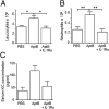
IL-1α–dependent inflammatory responses after i.p. injection of AptB. (A) Leukocytes (5.3 ± 0.6 x10⁶) were measured by flow cytometry after the injection of 30 × 106 AptB into the peritoneal cavity versus 3.5 ± 0.2 x 10⁶ in PBS condition. Injection of 30 mg/kg IL-1Ra before the injection of AptB induce significant reduction of leukocytes measured in peritoneal fluid (*P < 0.05). (B) Neutrophils (5.7 ± 0.6 x 10⁵) from data shown in A were measured by flow cytometry in peritoneal fluid after injection of AptB compared with 2.7 ± 0.5 x 10⁵ neutrophils in PBS condition and 2.2 ± 0.6 x 10⁵ in AptB and IL-1Ra condition (**P < 0.01). (C) KC concentrations (121 ± 20 pg/mL) obtained in serum 15 h after the i.p. injection of the AptB versus 39 ± 14 pg/mL in vehicule condition and 56.5 ± 21.2 in AptB + IL-1Ra condition (*P < 0.05; ANOVA test). Five independent experiments represent a total number of 6–11 mice for each condition.

Similar articles

Cited by

References

    1. Rock KL, Latz E, Ontiveros F, Kono H. The sterile inflammatory response. Annu Rev Immunol. 2010;28:321–342. - PMC - PubMed
    1. Zitvogel L, Kepp O, Kroemer G. Decoding cell death signals in inflammation and immunity. Cell. 2010;140:798–804. - PubMed
    1. Rider P, et al. IL-1alpha and IL-1beta recruit different myeloid cells and promote different stages of sterile inflammation. J Immunol. 2011;187:4835–4843. - PubMed
    1. Dinarello CA. Immunological and inflammatory functions of the interleukin-1 family. Annu Rev Immunol. 2009;27:519–550. - PubMed
    1. Kurt-Jones EA, Fiers W, Pober JS. Membrane interleukin 1 induction on human endothelial cells and dermal fibroblasts. J Immunol. 1987;139:2317–2324. - PubMed

Publication types

LinkOut - more resources