. 2011 Dec 20;108(51):20609-14.
doi: 10.1073/pnas.1118017108. Epub 2011 Dec 5.

Mouse digit tip regeneration is mediated by fate-restricted progenitor cells

Affiliations

Mouse digit tip regeneration is mediated by fate-restricted progenitor cells

Jessica A Lehoczky et al. Proc Natl Acad Sci U S A. .

Abstract

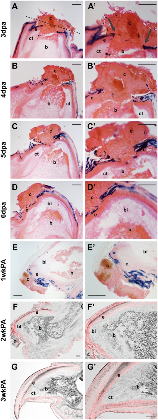
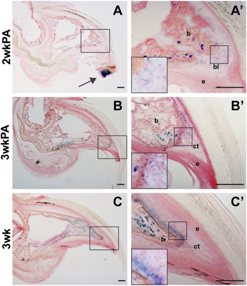
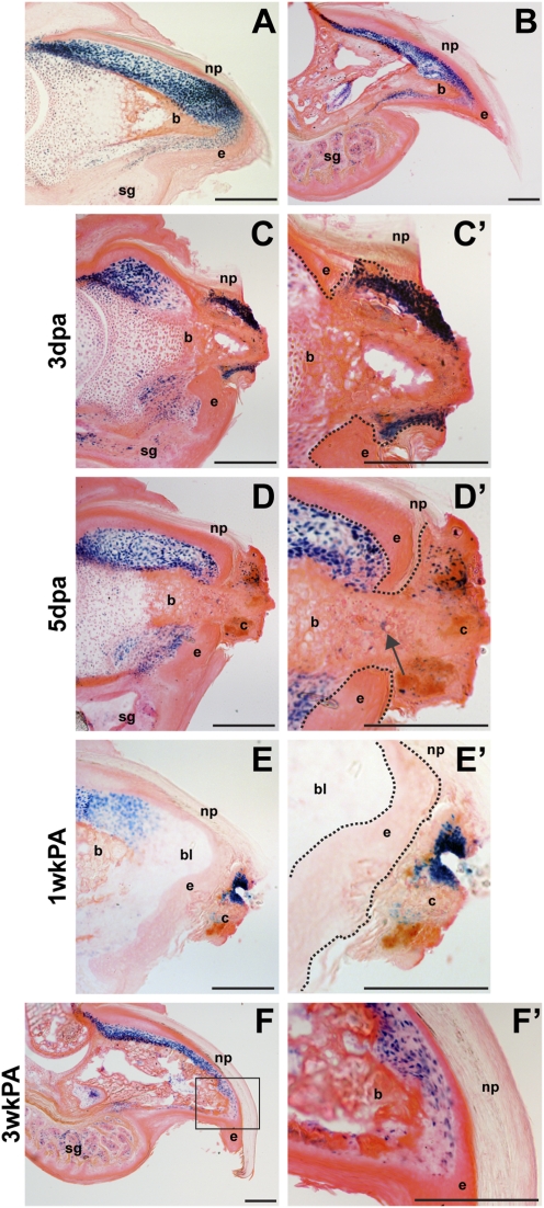
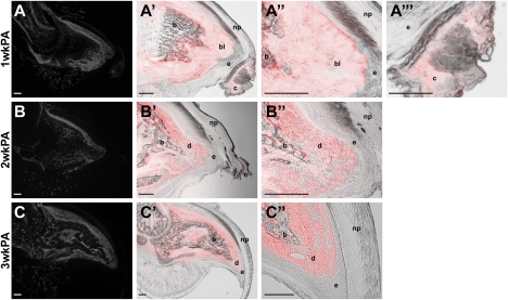
Regeneration of appendages is frequent among invertebrates as well as some vertebrates. However, in mammals this has been largely relegated to digit tip regeneration, as found in mice and humans. The regenerated structures are formed from a mound of undifferentiated cells called a blastema, found just below the site of amputation. The blastema ultimately gives rise to all of the tissues in the regenerate, excluding the epidermis, and has classically been thought of as a homogenous pool of pluripotent stem cells derived by dedifferentiation of stump tissue, although this has never been directly tested in the context of mammalian digit tip regeneration. Successful digit tip regeneration requires that the level of amputation be within the nail bed and depends on expression of Msx1. Because Msx1 is strongly expressed in the nail bed mesenchyme, it has been proposed that the Msx1-expressing cells represent a pluripotent cell population for the regenerating digit. In this report, we show that Msx1 is dynamically expressed during digit tip regeneration, and it does not mark a pluripotent stem cell population. Moreover, we show that both the ectoderm and mesoderm contain fate-restricted progenitor populations that work in concert to regenerate their own lineages within the digit tip, supporting the hypothesis that the blastema is a heterogeneous pool of progenitor cells.


Conflict of interest statement

The authors declare no conflict of interest.

Figures

Fig. 1.
Fig. 1.
Lineage of Krt14-expressing keratinocytes during digit tip regeneration. (A–E) X-gal stained (blue) Krt14-CreESR;R26R-lacZ digit sections from 3 dpa to 1 wkPA. (F and G) TdTomato (red) fluorescence overlaying differential interference contrast brightfield (grayscale) images of Krt14-CreESR1;R26R-CAG-tdTomato digit sections at 2 wkPA and 3 wkPA. Epidermal retraction and proximal attachment to terminal phalanx occurs between 1 and 3 dpa (A and A’, arrows). Dotted line shows approximate plane of amputation. Distally exposed bone becomes integrated into clot (B, B’, C, and C’) as epidermis covers the amputation site under the clot (C–E and C’–E’). Blastema formation occurs after epidermal closure (D and D’). bl, blastema; b, bone; c, clot; ct, connective tissue; e, epidermis. (Scale bars,100 μm.)
Fig. 2.
Fig. 2.
Lineage of Sp7-expressing osteoblasts during digit tip regeneration. X-gal stained Sp7-tTA-tetO-EGFP::Cre;R26R-lacZ digit sections. (A and A’) Images at 2 wkPA show descendants in clot (arrow), bone, and blastema, but not the epidermis. (B and B’) Images at 3 wkPA show descendants in bone and connective tissue but not epidermis. Staining resembles normal 3 wk nonregenerative digit osteoblast contribution (C and C’). Insets in A’–C’ show 40× magnification. bl, blastema; b, bone; ct, connective tissue; e, epidermis. (Scale bars, 100 μm.)
Fig. 3.
Fig. 3.
Expression of Msx1 during digit tip regeneration. Msx1-nlacZ x-gal stained digit sections during regeneration. PN3 (A) and 3 wk (B) unamputated digits show expression in dorsal and ventral dermis and throughout bone and sweat glands. (C and C’) Expression at 3 dpa is in normal stump domains and in the clot. (D and D’) Expression at 5 dpa is in the clot and a few bone cells where epidermis is closing under the clot (arrow). (E and E’) Expression at 1 wkPA remains in the clot but is absent from the blastema. (F and F’) Expression at 3 wkPA returns to a nonregenerative expression pattern and resembles B. Epidermis is outlined in higher-magnification pictures. bl, blastema; b, bone; c, clot; e, epidermis; np, nail plate; sg, sweat gland. (Scale bars, 200 μm.)
Fig. 4.
Fig. 4.
Lineage of Msx1-expressing cells during digit tip regeneration. TdTomato fluorescence from Msx1-CreERT2;R26R-CAG-tdTomato digit tips. (A–A’’’) Descendants at 1 wkPA are in blastema (A’’) and clot (A’’’) but not epidermis (A’–A’’’). (B–B’’) Descendants at 2 wkPA are found in bone and dermis but not epidermis. (C–C’’) Descendants at 3 wkPA have reestablished normal Msx1 expression pattern of the distal tip. (A–C) TdTomato expression with no overlay. (A’–A’’’, B’, B’’, C’, and C’’) Differential interference contrast brightfield images (grayscale) with tdTomato (red) fluorescent overlay. bl, blastema; b, bone; cl, clot; d, dermis; e, epidermis; np, nail plate. (Scale bar, 100 μm.)
Fig. 5.
Fig. 5.
Clot removal impairs bone regeneration. (A) Brightfield image of fibrin clot at 6 dpa. Inset: PN3 amputated digit tip, with dotted line depicting amputated tissue. (B–F) Alizerin red/alcian blue stained digit tip skeletons at 3 wkPA representing average extents of regeneration for the group. (B) Nonregenerate digit after nail removal. (C) Control digit after no clot removal. Digits after clot removal at 8 dpa (D), 7 dpa (E), and 6 dpa (F). (G) Boxplot analysis of clot removal experiment shows significant decrease in regeneration when the clot was removed at 6 dpa or 7 dpa, as measured by 2D area in pixels. Open circles represent 1.5 interquartile range outliers. Average nonregenerate digit tip area is denoted with a red line.

Similar articles

Cited by

References

    1. Thornton CS. The effect of apical cap removal on limb regeneration in Amblystoma larvae. J Exp Zool. 1957;134:357–381. - PubMed
    1. Mescher AL. Effects on adult newt limb regeneration of partial and complete skin flaps over the amputation surface. J Exp Zool. 1976;195:117–128. - PubMed
    1. Repesh LA, Oberpriller JC. Scanning electron microscopy of epidermal cell migration in wound healing during limb regeneration in the adult newt, Notophthalmus viridescens. Am J Anat. 1978;151:539–555. - PubMed
    1. Hay ED, Fischman DA. Origin of the blastema in regenerating limbs of the newt Triturus viridescens. An autoradiographic study using tritiated thymidine to follow cell proliferation and migration. Dev Biol. 1961;3:26–59. - PubMed
    1. Butler EG, O'Brien JP. Effects of localized x-radiation on regeneration of the urodele limb. Anat Rec. 1942;84:407–413.

Publication types

LinkOut - more resources